

Every minute, approximately one-fifth of your cardiac output passes through the kidneys via the renal arteries. The urinary system filters this blood, removes metabolic wastes such as urea, and regulates the body's water and salt balance to maintain homeostasis. This revision note covers the gross anatomy of the kidneys, ureters, bladder, and urethra; the microscopic functioning of the nephron during ultrafiltration and selective reabsorption; the homeostatic control of water balance by anti-diuretic hormone (ADH) via negative feedback; and medical applications including kidney dialysis and transplantation.
Excretion, Osmoregulation, and Macrostructure
Cells carry out thousands of chemical reactions that generate metabolic by-products. Excretion is the removal from the body of the waste products of metabolism. Do not confuse excretion with egestion, which is the removal of undigested food (faeces) from the alimentary canal. Because undigested food never enters body cells and does not take part in cellular metabolism, its elimination is not excretion.
The kidney is the central organ of the urinary system, but other human organs also carry out excretion:
| Organ | Waste Excreted |
|---|---|
| Kidneys | Urea, excess salts, and excess water (as urine) |
| Lungs | Carbon dioxide and water vapour |
| Skin | Water, mineral salts, and small amounts of urea (in sweat) |
| Liver | Bile pigments (produced from the breakdown of haemoglobin) |
Sweating primarily regulates body temperature; urea excretion in sweat is merely a minor secondary effect, and the kidneys remain the primary route for clearing urea produced by deamination of excess amino acids in the liver.
Core Functions of the Urinary System
- Excretion: Filtering out toxic nitrogenous waste (urea) along with excess water and mineral ions.
- Osmoregulation: The control of water and dissolved solute concentrations in the body fluids. This maintains homeostasis, ensuring that body cells neither swell and burst in a dilute environment nor shrivel in a concentrated one.
- pH Regulation: Helping to maintain blood pH within its narrow physiological range (around 7.4).
Macrostructure of the Urinary System
The system consists of four primary structures:
- Kidneys (two): Bean-shaped organs situated against the back wall of the upper abdomen, protected by the lower ribs. They filter blood, carry out selective reabsorption, and produce urine.
- Ureters (two): Muscular tubes extending from the renal pelvis of each kidney down to the bladder. They propel urine onward by waves of muscular contraction called peristalsis.
- Urinary Bladder (one): A hollow, distensible muscular organ in the pelvis that temporarily stores urine.
- Urethra (one): A single tube running from the neck of the bladder to the exterior of the body, carrying urine out during urination.
Urine release is controlled by sphincter muscle rings at the junction between the bladder and the urethra. Blood enters each kidney via a renal artery branching directly from the aorta, and cleaned blood leaves through a renal vein that empties into the inferior vena cava.
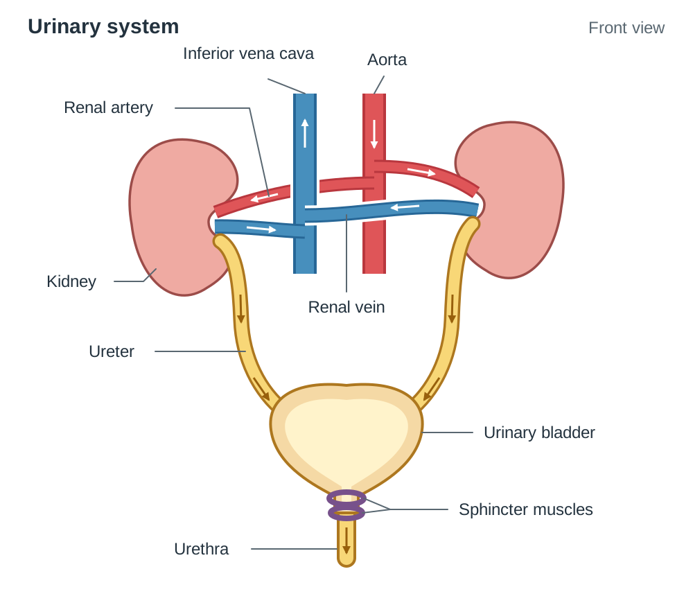
Labelled Diagram Checklist: Urinary System
In examinations, you may be asked to label a blank diagram or state the function of highlighted structures. Be prepared to identify:
- Kidney, renal artery, renal vein, ureter, urinary bladder, urethra, and sphincter muscle.
Internal Anatomy of the Kidney
A longitudinal cross-section of a kidney reveals three distinct regions:
- Renal Cortex: The outer reddish-brown layer. It contains the glomeruli, Bowman's capsule structures, and both the proximal and distal convoluted tubules. The cortex is the sole site of ultrafiltration and also carries out a large share of selective reabsorption.
- Renal Medulla: The darker, striated inner region divided into triangular renal pyramids. It contains the loops of Henle and the collecting ducts. The tissue fluid of the medulla is maintained at an exceptionally high salt concentration, which provides the osmotic pull required to reclaim water from collecting ducts.
- Renal Pelvis: The funnel-shaped white cavity at the centre of the kidney. Collecting ducts empty finished urine into the pelvis, which channels it directly into the ureter.
Note the distinction: reabsorption occurs in both the cortex and the medulla, but ultrafiltration occurs only in the cortex. The renal pelvis is where urine is collected, not where it is filtered or where solutes are reabsorbed.
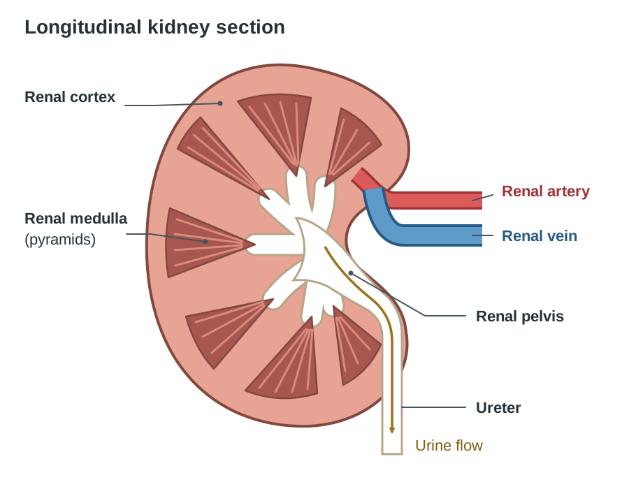
Labelled Diagram Checklist: Kidney Section
Be ready to identify and state the function of:
- Renal cortex, renal medulla (pyramids), renal pelvis, renal artery, renal vein, and ureter.
The Nephron: Architecture and Blood Supply
The nephron is the functional, microscopic unit of the kidney. Each human kidney contains over one million nephrons. The nephron works in two steps. Step one is deliberately wasteful: push almost everything small out of the blood, wastes and useful substances alike. Step two is careful: take back the useful substances and the water you need, and let the rest go as urine.
Blood Pathway Through the Nephron
The blood supply moves through two distinct capillary beds in series:
- Afferent arteriole: A branch of the renal artery with a relatively wide lumen that carries oxygenated, unfiltered blood into the glomerulus. A helpful mnemonic: Afferent Arrives, Efferent Exits.
- Glomerulus: A compact knot of capillaries nestled inside Bowman's capsule where filtration takes place.
- Efferent arteriole: The vessel carrying blood out of the glomerulus. Its lumen is noticeably narrower than that of the afferent arteriole.
- Peritubular capillaries: A dense network of capillaries branching from the efferent arteriole. They wrap closely around the tubules, so reabsorbed water and nutrients have only a very short distance to travel back into the blood.
Fluid Pathway Along the Nephron
Filtrate formed at the glomerulus travels through five connected regions before exiting as urine:
Collecting ducts receive fluid from several nephrons and run down through the medulla to empty into the renal pelvis.
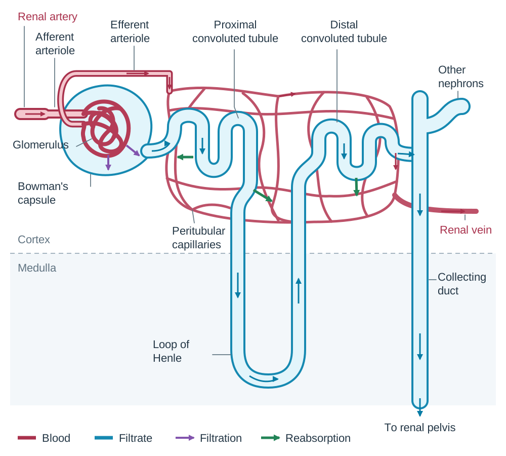
Labelled Diagram Checklist: Nephron
Be ready to label and provide one function for:
- Afferent arteriole, glomerulus, efferent arteriole, Bowman's capsule, proximal convoluted tubule, loop of Henle, distal convoluted tubule, collecting duct, and peritubular capillaries.
Stage 1: Ultrafiltration in Bowman's Capsule
Ultrafiltration occurs in the renal cortex between the glomerulus and the cup-shaped Bowman's capsule. Because the incoming afferent arteriole has a wider lumen than the outgoing efferent arteriole, blood encounters resistance as it exits the capillary knot. This structural bottleneck builds high hydrostatic pressure inside the glomerular capillaries.
This high pressure forces water and all small dissolved molecules through the porous capillary walls and across the basement membrane of Bowman's capsule into the tubule lumen:
- Substances filtered: Water, glucose, amino acids, urea, vitamins, and mineral salts pass into the capsule cavity to form glomerular filtrate.
- Substances retained: Red blood cells, white blood cells, platelets, and large plasma proteins (such as albumin and antibodies) are too large to pass through the filtration pores and remain inside the capillary blood.
Ultrafiltration is a non-selective, size-dependent physical process. It does not separate useful molecules from metabolic wastes. Small, valuable nutrients such as glucose and amino acids cross into the filtrate at the same concentration as they have in the blood plasma, exactly as wastes such as urea do — the filter cannot tell them apart.
The kidneys produce about 180 litres of glomerular filtrate every 24 hours. An adult body only holds about 35 to 40 litres of fluid in total, so if all of that filtrate left the body as urine you would be fatally dehydrated within hours. This is why the tubules must reabsorb almost all of it.
Stage 2: Selective Reabsorption Along the Tubules
Selective reabsorption recovers useful materials from the glomerular filtrate back into the surrounding peritubular capillaries. Approximately 99% of the filtered liquid volume returns to the blood as it moves through the nephron.
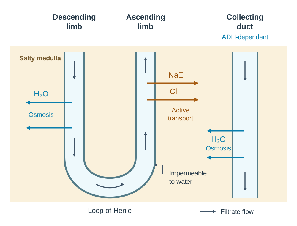
The Role of the Loop of Henle
The loop of Henle's job is to make the tissue fluid of the medulla very salty. Sodium and chloride ions are pumped out of the ascending limb, which is impermeable to water. That salty medulla creates a strong osmotic gradient that pulls water out of the collecting duct later on, which is why humans can produce urine that is more concentrated than their own blood.
| Nephron Region | Location | Substances Reabsorbed | Mechanism |
|---|---|---|---|
| Proximal Convoluted Tubule (PCT) | Cortex | 100% of glucose and amino acids; most mineral salts; ~65% of water | Active transport (glucose, amino acids, salts), diffusion, and osmosis (water) |
| Descending Limb of Loop of Henle | Medulla | Water only (permeable to water, impermeable to salts) | Osmosis into the concentrated tissue fluid of the medulla |
| Ascending Limb of Loop of Henle | Medulla | Sodium and chloride ions (impermeable to water) | Sodium and chloride ions are removed from the filtrate into the medulla, mainly by active transport. This limb is impermeable to water, which is what makes the medulla tissue fluid so salty |
| Distal Convoluted Tubule (DCT) | Cortex | Selected salts and some water; blood pH adjustments | Active transport (salts) and osmosis (water) |
| Collecting Duct | Medulla | Water only | Osmosis into the salty medulla; permeability controlled by ADH |
Structural Adaptations of the Proximal Convoluted Tubule
The PCT reabsorbs all filtered food molecules and the majority of water. It displays four vital cellular adaptations:
- Microvilli: The internal epithelial lining has millions of microscopic folds, vastly expanding the surface area for membrane transport proteins.
- Abundant mitochondria: Cells contain large numbers of mitochondria to supply ATP for the active transport of glucose, amino acids, and sodium ions against concentration gradients.
- Thin wall: The wall is one cell thick, providing a short diffusion distance into the adjacent blood supply.
- Dense capillary network: Peritubular capillaries wrap closely around the PCT and carry the reabsorbed substances away in the blood, keeping the concentration gradient for reabsorption steep.
The fluid leaving the distal convoluted tubule is still being adjusted. Once its final water content has been set in the collecting duct under the control of ADH, the fluid is called urine: water, urea and excess salts.
Composition of Urine and Diagnostic Urine Analysis
Comparing solute concentrations across blood plasma, glomerular filtrate, and urine demonstrates the precision of nephron function (values given in ):
| Substance | Blood Plasma | Glomerular Filtrate | Normal Urine | Biological Explanation |
|---|---|---|---|---|
| Protein | 8.0 | 0.0 | 0.0 | Plasma proteins are too large to pass through the glomerular capillary pores and basement membrane during ultrafiltration. |
| Glucose | 0.1 | 0.1 | 0.0 | Glucose filters freely into Bowman's capsule but is 100% reabsorbed in the PCT by active transport. |
| Urea | 0.03 | 0.03 | 2.0 | Urea filters freely and is not reabsorbed in significant amounts. As ~99% of filtered water is reclaimed, the remaining urea becomes highly concentrated. |
| Mineral Salts | 0.9 | 0.9 | 1.5 | Salts filter freely. Most are selectively reabsorbed to maintain osmotic balance, while the excess is excreted in a smaller water volume. |
Normal Urine Composition
Normal urine is approximately 95% water. The dissolved solutes consist primarily of urea, along with mineral salts (chiefly sodium chloride) and small amounts of uric acid and creatinine. Normal urine contains no glucose, no protein, and no blood cells.
Interpreting an Abnormal Urine Sample
- Glucose present: When blood glucose concentration exceeds the capacity of the active transport carrier proteins in the PCT, the excess glucose cannot be reabsorbed and passes into urine. This is a primary diagnostic indicator of untreated diabetes mellitus.
- Protein or red blood cells present: Indicates damage to the glomerular capillaries or the basement membrane of Bowman's capsule. Molecules and cells that are normally too large to filter are leaking into the filtrate, showing that the kidney's filtration barrier has failed.
Diagnostic rule of thumb: in ultrafiltration the barrier decides what leaves the blood, while in selective reabsorption the tubule decides what returns. Therefore, protein in urine points to a filtration fault, whereas glucose in urine points to a reabsorption capacity being overwhelmed.
Osmoregulation and the Action of ADH
Diuresis means the production of a large volume of urine. An anti-diuretic hormone works against this: it reduces the volume of urine produced by causing more water to be reabsorbed. Anti-diuretic hormone (ADH) is produced by neurosecretory cells in the hypothalamus and stored in and released from the posterior pituitary gland. ADH targets the cells of the distal convoluted tubule and the collecting duct, increasing their permeability to water.
The Six-Line ADH Exam Marking Skeleton
For any long-form question on ADH, structure your answer using these six steps:
- Blood plasma becomes too concentrated (dehydration) OR too dilute (high water intake).
- Osmoreceptors in the hypothalamus detect this change.
- The posterior pituitary gland releases more ADH (if concentrated) OR less ADH (if dilute).
- ADH travels in the bloodstream to make the DCT and collecting duct more permeable (or less permeable) to water.
- More water is reabsorbed by osmosis into the blood (or less water is reabsorbed).
- The kidneys produce a small volume of concentrated urine (or a large volume of dilute urine).
Case 1: Dehydration, Heavy Sweating, or High Salt Intake
- Water loss causes blood plasma to become hypertonic (solute concentration rises).
- Hypothalamic osmoreceptors detect the increased osmotic pressure of the blood.
- The posterior pituitary releases more ADH into the circulation.
- ADH binds to receptors on the DCT and collecting duct, opening water channels and making the tubule walls more permeable to water.
- More water leaves the filtrate by osmosis, moving into the salty medulla and returning to the bloodstream via peritubular capillaries.
- The individual excretes a small volume of concentrated, dark urine.
- Blood solute concentration returns to the normal set point, which switches off the stimulus for ADH secretion (negative feedback).
Case 2: Large Fluid Intake or Low Salt Diet
- Ingestion of water causes blood plasma to become hypotonic (solute concentration falls).
- Hypothalamic osmoreceptors detect the drop in osmotic pressure.
- The posterior pituitary releases less ADH (or stops secretion).
- The DCT and collecting duct remain impermeable to water.
- Very little water is reabsorbed from these sections, keeping water in the tubule lumen.
- The individual excretes a large volume of dilute, pale urine.
- Solute concentration in the blood rises back to normal set levels.
Kidney Failure, Dialysis, and Scientific Models
Kidney failure occurs when the kidneys can no longer filter metabolic waste or regulate water and electrolyte balance. Urea, excess salts, and fluid accumulate in the blood, leading to a toxic condition that is fatal if left untreated.
Treatments for Kidney Failure
- Haemodialysis (Kidney Dialysis): The patient's blood is diverted through a dialysis machine where it passes along one side of a selectively permeable membrane, while dialysis fluid flows in the opposite direction on the other side. Urea and excess mineral ions diffuse out of the blood, down their concentration gradients, into the dialysis fluid. Blood cells and plasma proteins are too large to cross the membrane pores and remain in the patient's blood. The dialysis fluid contains glucose and essential mineral salts at normal blood concentrations, preventing any loss of essential nutrients. Dialysis must be carried out several times per week.
- Kidney Transplant: A healthy donor kidney is surgically connected to the recipient's iliac blood vessels and bladder. While a successful transplant restores continuous, normal filtration and frees the patient from dialysis, the patient must take immunosuppressive drugs for life to prevent organ rejection.
Scientific Models: Dialysis Tubing and Ultrafiltration
In laboratory science, models simplify complex biological phenomena. Ultrafiltration can be modelled using Visking (dialysis) tubing containing a solution of starch, glucose, and protein submerged in water:
- What it models: The microscopic pores of the tubing permit small dissolved molecules (glucose and water) to diffuse out, while retaining large molecules (starch and protein). This represents the size-selective action of the glomerular basement membrane.
- Limitations of the model: Dialysis tubing relies entirely on passive diffusion across an unpowered membrane. It lacks the hydrostatic blood pressure that drives living filtration, cannot perform active transport, and cannot respond to hormones like ADH.
- Technology link: A clinical kidney dialysis machine is this exact model scaled up and engineered as medical technology.
Evaluating Urinary Output Data
When interpreting experimental data regarding urine production:
- Concentration versus total excretion: A dark, concentrated urine sample does not mean more urea was eliminated; it usually means more water was reabsorbed. Total waste excretion can only be determined by multiplying solute concentration by total urine volume over time.
- Fair test controls: When comparing urine output under different experimental conditions, variables such as ambient temperature, physical exertion, prior hydration status, and dietary salt intake must be kept strictly constant.
Key terms
- Excretion
- The removal from the body of the waste products of metabolism.
- Osmoregulation
- The control of the water and dissolved solute concentrations in the body fluids of an organism.
- Homeostasis
- The maintenance of a constant internal environment in an organism.
- Deamination
- The breakdown of excess amino acids in the liver to form urea.
- Nephron
- The microscopic structural and functional unit of the kidney that filters blood and produces urine.
- Cortex (of kidney)
- The outer region of the kidney containing glomeruli, Bowman's capsules, and convoluted tubules; the site of ultrafiltration and major selective reabsorption.
- Medulla (of kidney)
- The inner region of the kidney containing the loops of Henle and collecting ducts, characterised by salty tissue fluid that drives water reabsorption.
- Renal Pelvis
- The central funnel-shaped cavity of the kidney that collects urine from the collecting ducts and channels it into the ureter.
- Glomerulus
- A dense knot of high-pressure capillaries inside Bowman's capsule where ultrafiltration of blood occurs.
- Bowman's Capsule
- The cup-like sac at the beginning of the nephron that encloses the glomerulus and collects glomerular filtrate.
- Glomerular Filtrate
- The fluid forced out of glomerular capillaries into Bowman's capsule, containing water, glucose, amino acids, salts, and urea, but lacking blood cells and large proteins.
- Ultrafiltration
- The filtration of blood under high hydrostatic pressure across the glomerular capillary walls into Bowman's capsule.
- Selective Reabsorption
- The recovery of useful substances (such as glucose, amino acids, salts, and water) from the filtrate back into the blood of peritubular capillaries.
- Anti-Diuretic Hormone (ADH)
- A hormone produced by the hypothalamus and released by the posterior pituitary that increases the water permeability of the distal convoluted tubule and collecting duct.
- Diuresis
- The production of a large volume of dilute urine.
- Negative Feedback
- A control mechanism in which a change away from a normal set point triggers a physiological response that counteracts the initial change.
Check yourself
What structural difference between the afferent and efferent arterioles generates high blood pressure in the glomerulus?
The afferent arteriole has a wider lumen than the narrower efferent arteriole, creating a pressure bottleneck in the glomerular capillaries.
Distinguish between excretion and egestion, giving one example of a substance removed by each.
Excretion is the removal of metabolic waste products produced by body cells (e.g. urea or carbon dioxide), whereas egestion is the removal of undigested dietary material that was never absorbed into cells (e.g. dietary fibre / faeces).
A patient's urine sample contains 0.4 g / 100 cm³ of protein. Suggest what this indicates and explain your reasoning.
This indicates damage to the glomerulus or Bowman's capsule basement membrane. Plasma proteins are normally too large to pass through the filtration pores during ultrafiltration; their presence in urine shows that the kidney's filtration barrier has failed.
Name three structural adaptations of the proximal convoluted tubule that facilitate selective reabsorption.
Microvilli lining the lumen to provide a large surface area; numerous mitochondria to supply ATP for active transport; and a thin wall (one cell thick) for short diffusion distance.
What is the primary function of the loop of Henle in the kidney medulla?
To actively pump sodium and chloride ions into the tissue fluid of the medulla, creating a salty, concentrated environment that draws water out of the collecting duct by osmosis.
Why does a clinical kidney dialysis machine use dialysis fluid containing normal concentrations of glucose and mineral salts?
So that there is no concentration gradient between the patient's blood and the dialysis fluid for these essential nutrients, preventing glucose and vital salts from diffusing out of the blood.