

Semiconservative DNA Replication and the Cell Cycle
DNA replication occurs during interphase of the cell cycle, before nuclear division begins. Replication is essential so that each chromosome is duplicated into two identical sister chromatids joined at the centromere. During mitosis, these chromatids are separated into opposite daughter cells. Without prior replication, each new cell would receive only half the necessary genetic instructions. Furthermore, any copying mistakes made during replication become permanent and are passed to all descendant cells.
Replication follows a semiconservative mechanism because each newly formed double helix conserves one original parent strand and incorporates one newly made daughter strand.
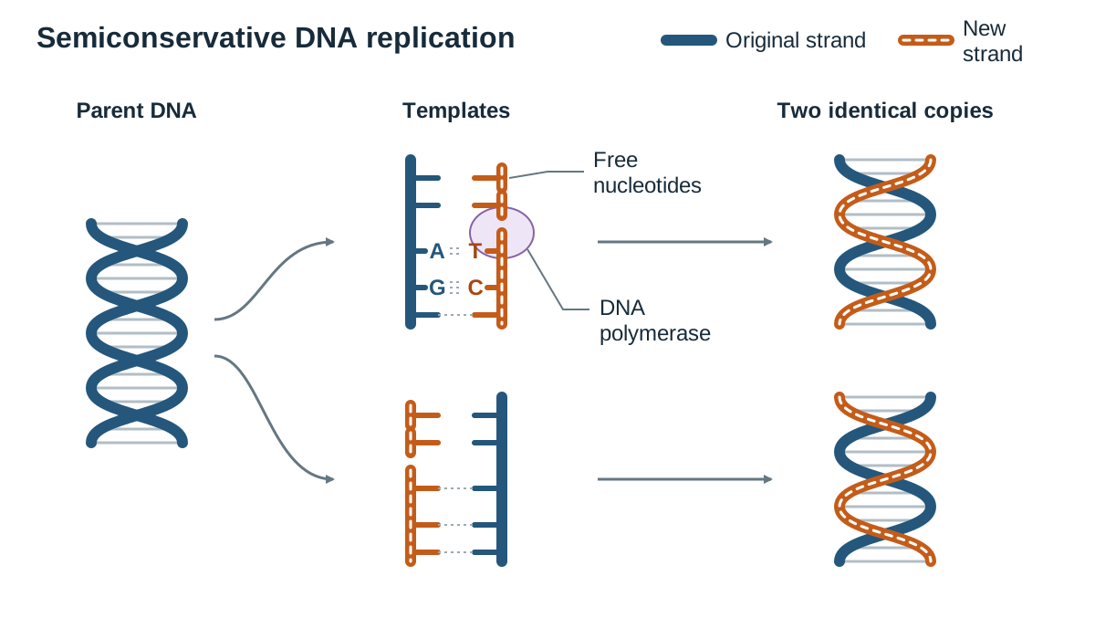
Steps in DNA Replication
- Enzymes break the hydrogen bonds between base pairs, unzipping the parent double helix and exposing the bases on both template strands.
- Free DNA nucleotides present in the nucleoplasm move to the exposed template strands and align opposite their complementary bases ( with , and with ).
- The enzyme DNA polymerase joins adjacent nucleotides by building the covalent sugar-phosphate backbone of the new strand. DNA polymerase also proofreads the newly added bases, correcting mismatched pairs.
- Each new double-stranded molecule rewinds into a double helix, producing two genetically identical copies of the original DNA.
Scientific Models of DNA Replication
Biologists use diagrams and physical models to represent replication. A typical diagram shows the parent double helix partially unzipped into a 'Y-shaped' replication fork, with free nucleotides aligning along each template strand and DNA polymerase moving behind them, resulting in two shaded strands (one old, one new). While useful, these models have limitations:
- They are drawn flat like a ladder, ignoring the natural three-dimensional twist of the double helix.
- They show only a handful of bases, whereas real human chromosomes contain millions of base pairs.
- They depict replication as a static, step-by-step event, whereas in living cells it is a continuous, rapid biochemical process involving many coordinating enzymes.
Transcription: Synthesising Messenger RNA
Transcription is the first stage of protein synthesis. It takes place in the nucleus, where the genetic code of a specific gene on DNA is transcribed into a single strand of messenger RNA (mRNA).
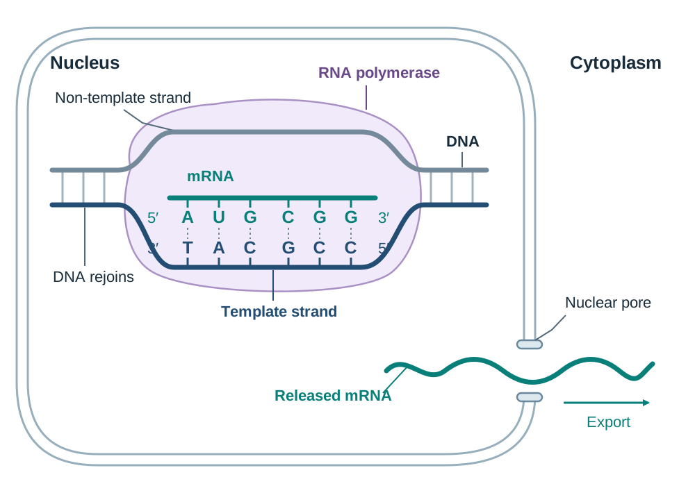
Mechanism of Transcription
- The enzyme RNA polymerase binds to the DNA at the start of a gene (a specific base sequence called a promoter).
- RNA polymerase unwinds the double helix and breaks the hydrogen bonds between complementary bases, exposing the template strand.
- Free RNA nucleotides present in the nucleoplasm align opposite their complementary bases on the DNA template strand: cytosine pairs with guanine, guanine pairs with cytosine, thymine on DNA pairs with adenine on RNA, and adenine on DNA pairs with uracil on RNA.
- RNA polymerase joins the RNA nucleotides together to form the continuous single strand of mRNA.
- When RNA polymerase reaches a termination signal at the end of the gene, the newly formed mRNA transcript detaches.
- The DNA template strand re-bonds with its partner strand and rewinds into a double helix. The mRNA transcript leaves the nucleus through a nuclear pore and enters the cytoplasm.
Translation: Assembling the Polypeptide Chain
Translation converts the codon sequence of an mRNA molecule into a specific sequence of amino acids joined by peptide bonds. This process takes place at ribosomes in the cytoplasm.
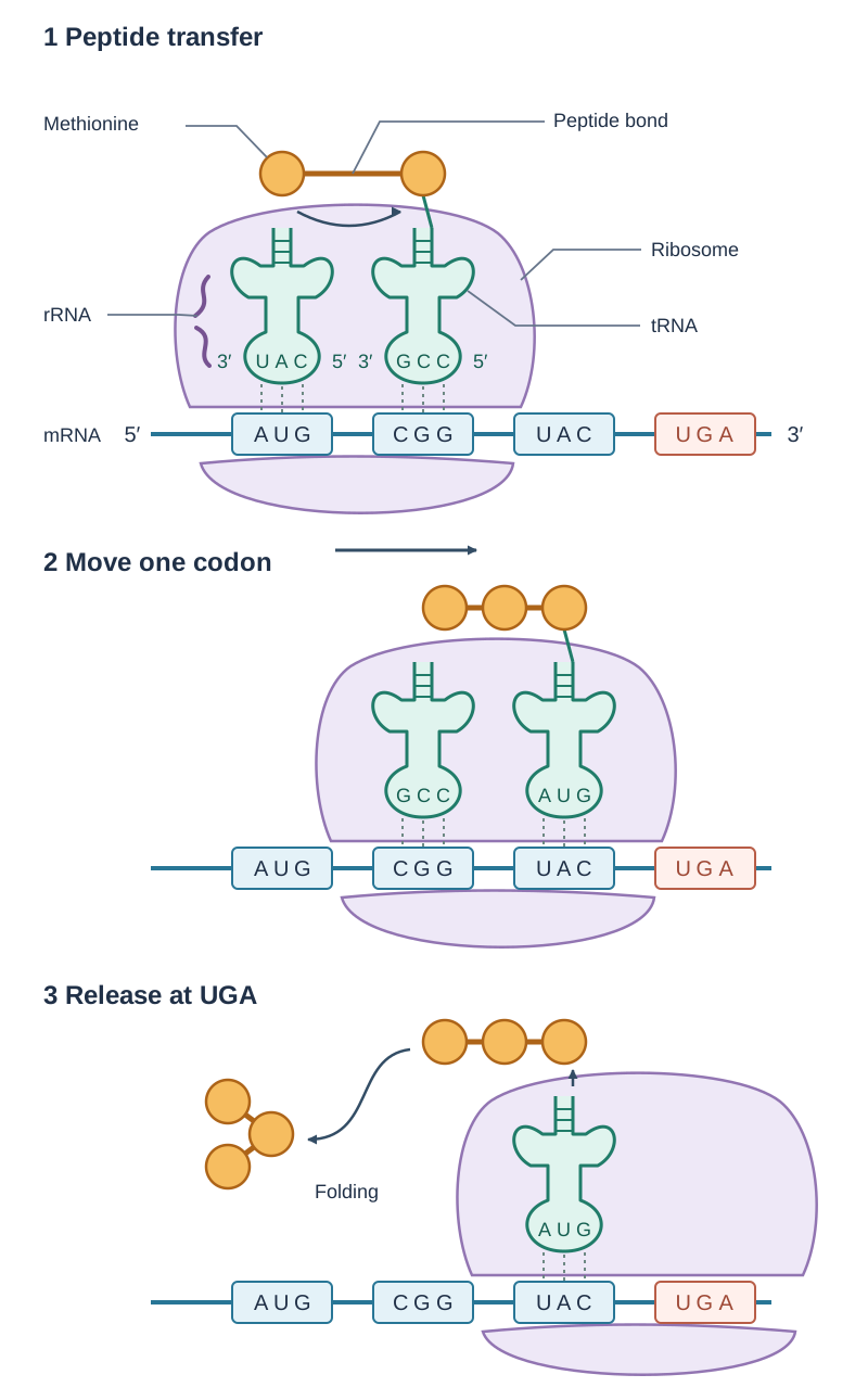
Steps in Translation
- Initiation: The mRNA strand docks onto a ribosome. The ribosome reads the strand until it locates the start codon (AUG). An initiator tRNA carrying the amino acid methionine binds to the start codon using its complementary anticodon (UAC).
- Elongation: The ribosome moves along the mRNA, reading one codon at a time. A second tRNA carrying its matching amino acid binds to the adjacent codon. The ribosome (its rRNA) catalyses the formation of a peptide bond between neighbouring amino acids. Once the bond forms, the first tRNA releases its amino acid and leaves the ribosome to collect another free amino acid in the cytoplasm. The ribosome then steps forward by one codon.
- Termination and Folding: Elongation continues until the ribosome reaches one of the three stop codons: UAA, UAG, or UGA. A stop codon does not code for an amino acid, and no tRNA molecule possesses a matching anticodon. Translation ceases, the completed polypeptide chain detaches from the ribosome, and it folds into its functional three-dimensional shape (such as a fibrous structural protein or a globular enzyme).
Modelling Translation
A standard model of translation shows the mRNA passing between ribosomal subunits, labelled codons grouped in threes, tRNAs docking at adjacent sites, and a growing chain of amino acid circles connected by peptide bonds. Like replication models, this diagram simplifies a complex system: it omits the true 3D shape of ribosomal subunits and shows translation as stationary frames rather than a continuous, rapid process adding several amino acids per second.
Roles of RNA Types in the Cell
Three distinct forms of RNA coordinate protein synthesis. The specification asks you to know the role of mRNA, tRNA and rRNA in protein synthesis. For each, learn its full name, where it is found and its job:
| RNA Type | Full Name | Cellular Location | Role in Protein Synthesis |
|---|---|---|---|
| mRNA | Messenger RNA | Synthesised in the nucleus; travels to the cytoplasm and binds to ribosomes | Carries the transcribed genetic instructions from nuclear DNA to the ribosome in groups of three bases called codons. |
| tRNA | Transfer RNA | Free in the cytoplasm; docks temporarily at the ribosome | Transports a specific amino acid to the ribosome and matches its anticodon to the complementary mRNA codon. |
| rRNA | Ribosomal RNA | Assembled in the nucleolus; forms the physical structure of ribosomes in the cytoplasm | Makes up the structural framework of the ribosome and catalyses peptide bond formation between amino acids. |