

Human reproduction is sexual: a sperm from the father and an egg from the mother join together, and the single cell that results grows into a baby inside the mother's uterus. Each parent's body cells have 46 chromosomes, but the sperm and egg have only 23 each, so when they fuse the normal 46 is restored. This topic follows that story in order: the two reproductive systems, how the gametes are made, the monthly cycle in the female, pregnancy and birth, and finally the hormones that control it all.
Male Reproductive System: Anatomy, Spermatogenesis, and Endocrine Control
The male reproductive system produces haploid sperm, synthesises sex hormones, and delivers semen into the female reproductive tract.
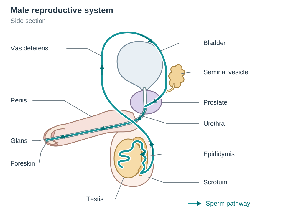
Structure and Function of Male Organs
- Testes: the two male gonads (sex organs), which hang outside the body in the scrotum. They contain thousands of tightly packed seminiferous tubules lined with diploid germ cells that divide by meiosis to produce haploid sperm cells. Interstitial cells between the tubules synthesise testosterone.
- Scrotum: A sac of skin holding the testes outside the abdominal cavity. It keeps testicular temperature at roughly 35°C (2°C below core body temperature of 37°C), which is the optimum temperature for meiosis and viable sperm production.
- Epididymis: A coiled tube resting along the posterior surface of each testis. Immature sperm pass here to mature, develop motility, and be stored until ejaculation. Unused sperm are reabsorbed.
- Vas Deferens (Sperm Duct): Muscular tubes leading from each epididymis into the pelvic cavity to join the urethra, propelling sperm forward by peristalsis during ejaculation.
- Seminal Vesicles and Prostate Gland: Accessory glands that produce seminal fluid. The seminal vesicles secrete a fluid rich in fructose to nourish sperm respiration. The prostate gland adds an alkaline fluid that neutralises acidity in the vagina and activates sperm. Seminal fluid combined with sperm cells forms semen.
- Urethra: A duct running through the penis that carries urine from the bladder or semen during ejaculation, though never at the same time due to an involuntary sphincter.
- Penis, Glans, and Foreskin: The external organ that introduces sperm into the female tract. It contains erectile tissue that engorges with blood during arousal. The sensitive tip is the glans, covered by a protective fold of skin called the foreskin.
The Pathway of Sperm
Trace the route of sperm in order: seminiferous tubules (testes) → epididymis → vas deferens → urethra → out through the penis. Diagram practice: Cover the labels on a diagram of the male reproductive system and practice naming these parts in the order sperm travel through them.
Structure of a Mature Sperm
A mature sperm is a microscopic, motile cell with three regions:
- Head: Contains the haploid nucleus (23 chromosomes) capped by an acrosome, an enzyme-filled sac that digests through the outer layers of the egg.
- Midpiece: Packed with mitochondria that carry out aerobic respiration to generate ATP for swimming.
- Tail (Flagellum): A whip-like structure that propels the sperm forward.
Hormonal Control in Males
At puberty, the anterior pituitary gland secretes two gonadotropic hormones:
- FSH (Follicle-Stimulating Hormone): Stimulates germ cells in the seminiferous tubules to divide by meiosis, initiating and sustaining continuous sperm production.
- LH (Luteinising Hormone): Stimulates the interstitial cells of the testes to secrete testosterone.
- Testosterone: Triggers the development of primary sex organs and secondary sexual characteristics at puberty (voice deepening, facial and body hair, broader shoulders, and muscle development), while maintaining spermatogenesis through life.
Female Reproductive System, Gametogenesis, and Fertilisation
The female reproductive system produces haploid ova, synthesises ovarian hormones, provides the site for fertilisation, and sustains the developing child throughout gestation.
Structure and Function of Female Organs
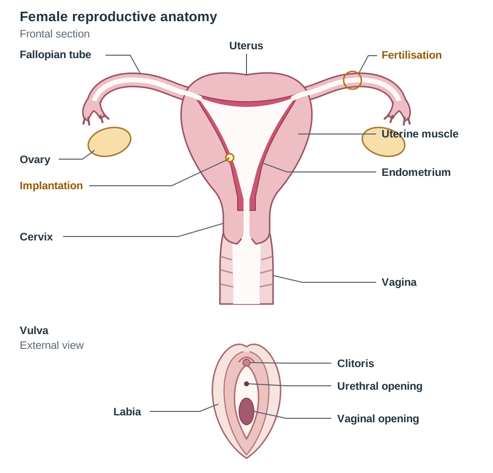
- Ovaries: The two female gonads located in the pelvic cavity. They produce haploid eggs by meiosis within cellular follicles and secrete oestrogen and progesterone.
- Fallopian Tubes (Oviducts): Muscular tubes lined with cilia extending from near each ovary to the uterus. The funnel-shaped end captures the ovulated egg, and cilia together with peristalsis move the egg toward the uterus. Fertilisation normally occurs in the fallopian tube.
- Uterus (Womb): A pear-shaped, thick-walled muscular organ. Its outer wall consists of smooth muscle that contracts during labour, while its vascular, glandular inner lining is the endometrium, where the embryo implants.
- Cervix: The muscular neck at the base of the uterus that opens into the vagina. It produces mucus to regulate sperm entry and dilates during labour.
- Vagina: An elastic muscular tube extending from the cervix to the exterior. It receives semen during intercourse and acts as the birth canal during labour.
- Vulva, Labia, and Clitoris: The external genitalia are collectively called the vulva. The labia are folds of skin protecting the vaginal opening. The clitoris sits anterior to the urethral opening and contains sensitive nerve endings and erectile tissue.
Diagram practice: Cover the labels on a female system diagram and name the structures in the sequence an egg travels: ovary → fallopian tube → uterus → cervix → vagina.
Structure of a Mature Egg (Ovum)
- Nucleus: Haploid, containing 23 chromosomes.
- Cytoplasm: Large volume containing food reserves that nourish the embryo during its first days before implantation.
- Cell membrane: Once a sperm enters, this membrane changes into a fertilisation membrane that prevents polyspermy (entry of any further sperm).
- Outer coats: A jelly-like layer surrounded by follicle cells. Digestive enzymes from the sperm acrosome break through these layers.
Comparing Sperm and Egg
- Sperm: Tiny, motile (has a flagellum and mitochondria), produced continuously from puberty in vast numbers.
- Egg: Much larger, non-motile, containing abundant cytoplasm and food reserves, with usually only one released per 28-day cycle.
The Biological Process of Fertilisation
Fertilisation is the fusion of the haploid nucleus of a sperm with the haploid nucleus of an egg to form a diploid zygote.
- Sperm swim through the cervix and uterus into the fallopian tube.
- The acrosome of the sperm releases enzymes that digest a path through the outer protective layers of the egg.
- The sperm head fuses with the egg membrane. The membrane immediately changes into a fertilisation membrane, blocking all other sperm.
- The haploid sperm nucleus () fuses with the haploid egg nucleus () to form a single diploid zygote ().
The Menstrual Cycle and Hormonal Feedback Loops
The menstrual cycle is a repeating cycle of approximately 28 days that prepares the endometrium for potential pregnancy. It is regulated by two pituitary hormones (FSH and LH) and two ovarian hormones (oestrogen and progesterone).
The four phases are not four separate blocks of time — the follicular phase begins on Day 1, so it overlaps with menstruation.
| Phase | Cycle Days | Ovarian Activity | Uterine Endometrium | Dominant Hormones |
|---|---|---|---|---|
| Menstruation | Days 1–5 | The previous corpus luteum degenerates; a new follicle begins to develop under rising FSH. | Without progesterone, the functional layer of the endometrium breaks down and sheds with blood through the vagina. | Oestrogen and progesterone are low; FSH begins to rise. |
| Follicular phase | Days 1–14 (overlaps menstruation) | FSH stimulates a follicle to develop into a mature Graafian follicle, which secretes oestrogen. | From Day 5 onwards, oestrogen repairs and thickens the endometrium with new blood vessels. | Rising oestrogen, which exerts negative feedback on FSH to stop extra follicles maturing. |
| Ovulation | Day 14 | The Graafian follicle ruptures through the ovarian surface, releasing the egg into the fallopian tube. | The endometrium continues to thicken. | High oestrogen triggers a sharp surge in LH, directly causing ovulation. |
| Luteal phase | Days 15–28 | LH transforms the empty Graafian follicle into the corpus luteum, which secretes progesterone. | Progesterone makes the endometrium thick, glandular, and vascular, ready for implantation. | High progesterone and moderate oestrogen. Progesterone inhibits FSH and LH by negative feedback. |
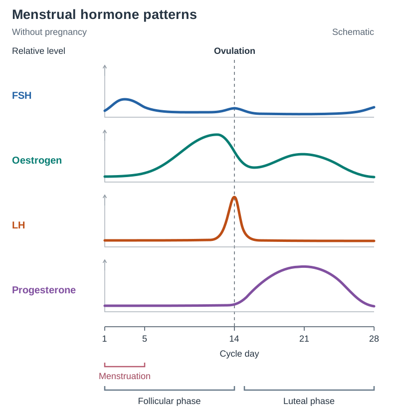
Reading the Hormone Graph
Exam questions frequently present a graph of hormone levels plotted against days:
- FSH: Low baseline with a small rise early in the cycle (Days 1–5) to stimulate follicle development.
- Oestrogen: Rises steadily during the follicular phase and peaks sharply just before Day 14.
- LH: Stays low until a sharp, distinct spike at Day 14. This spike is the direct signal for ovulation.
- Progesterone: Stays low until Day 14, climbs to a broad peak around Day 21 as the corpus luteum functions, then drops sharply by Day 28 if pregnancy does not occur.
Post-Luteal Outcomes
- If fertilisation does not occur: High progesterone suppresses LH. Deprived of LH, the corpus luteum degenerates by Day 28. Progesterone and oestrogen levels plummet, causing the endometrium to break down (menstruation). This drop relieves negative feedback, allowing pituitary FSH to rise again.
- If fertilisation occurs: The implanting blastocyst secretes hCG (human chorionic gonadotrophin). This hormone maintains the corpus luteum for the first 10 to 12 weeks of pregnancy, keeping progesterone high so the endometrium is not shed.
Pregnancy: From Zygote to Birth, Germ Layers, and the Placenta
Human gestation lasts roughly 38 weeks (266 days) from fertilisation. Early development follows a strict chronological order:
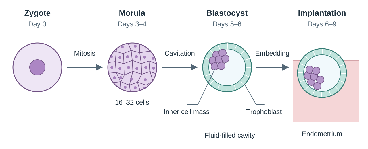
Early Developmental Stages
- Zygote: The single diploid cell formed by fertilisation in the upper fallopian tube.
- Cleavage: Rapid mitotic cell divisions of the zygote without an overall increase in size as it moves toward the uterus.
- Morula: A solid ball of 16 to 32 undifferentiated cells formed around Days 3 to 4.
- Blastocyst: A hollow, fluid-filled sphere formed around Days 5 to 6, containing two parts:
- Trophoblast: The outer single layer of cells that invades the endometrium and forms the embryonic part of the placenta.
- Inner cell mass: The inner group of cells that develops into the embryo proper.
- Implantation: The process where the blastocyst attaches and embeds into the vascular endometrium around Days 6 to 9.
Primary Germ Layers
Following implantation, the inner cell mass differentiates into three primary germ layers:
- Ectoderm: Gives rise to the nervous system (brain and spinal cord) and the epidermis of the skin.
- Mesoderm: Gives rise to skeletal muscle, bone, blood, and the circulatory system (heart and blood vessels).
- Endoderm: Gives rise to the inner lining of the alimentary canal and respiratory system, as well as the liver and pancreas.
The Amnion and Amniotic Fluid
The developing embryo is enclosed by a thin membrane called the amnion, which secretes amniotic fluid. Functions of amniotic fluid:
- Cushions the foetus against physical shock and mechanical injury.
- Keeps temperature around the foetus constant.
- Prevents the foetus from drying out and prevents foetal skin adhering to membranes.
- Allows the foetus to move freely, aiding muscular and skeletal development.
- Lubricates the birth canal when the amnion ruptures during labour.
The Placenta and Umbilical Cord
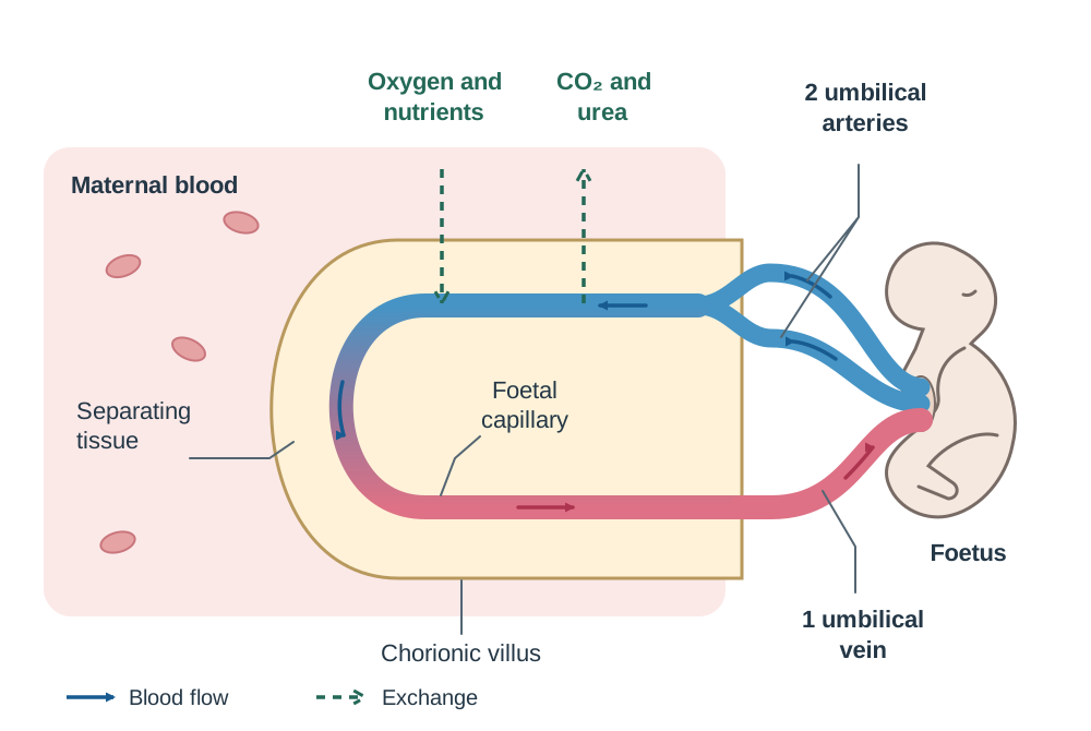
The placenta is formed from embryonic tissue (chorionic villi from the trophoblast) and maternal tissue (endometrium). The foetus connects to it via the umbilical cord, which contains two umbilical arteries (carrying deoxygenated blood and wastes from foetus to placenta) and one umbilical vein (carrying oxygen and nutrients from placenta to foetus).
- Two biological reasons why maternal and foetal blood must not mix:
- Maternal blood pressure in arteries is high enough that direct entry would rupture fragile foetal capillaries.
- Incompatible blood groups (such as ABO or Rhesus systems) would cause the mother's antibodies to clump together (agglutinate) or destroy the foetus's red blood cells, which can be fatal.
- Exchange function: Oxygen, glucose, amino acids, and antibodies cross from mother to foetus by diffusion and active transport. Carbon dioxide and urea cross from foetus to mother. Harmful substances such as alcohol, nicotine, and certain viruses can also cross.
- Endocrine function: The corpus luteum secretes progesterone for the first 10 to 12 weeks. After that, the fully developed placenta takes over progesterone synthesis, maintaining the endometrium and keeping the myometrium relaxed until birth.
Parturition (Birth)
From fertilisation to the end of week 8, the developing human is an embryo (organogenesis). From week 9 until birth, it is a foetus (growth and tissue maturation). Birth occurs in three stages:
- Stage 1 (Dilation): Placental progesterone falls and oxytocin causes rhythmic contractions of the uterine muscle. The cervix dilates to 10 cm and the amnion ruptures ('waters breaking').
- Stage 2 (Delivery): Strong uterine contractions push the baby through the cervix and vagina, usually head first. The umbilical cord is clamped and severed.
- Stage 3 (Afterbirth): Continued contractions detach and expel the placenta and foetal membranes within 5 to 30 minutes.
Hormonal Modelling Across the Human Lifespan (Higher Level)
At Higher Level, you must model the integrated actions of seven reproductive hormones across the human lifespan using gland origins, target organs, and feedback mechanisms.
Summary of Reproductive Hormones
| Hormone | Site of Production | Primary Target Organ | Main Effects |
|---|---|---|---|
| Testosterone | Interstitial cells of testes | Body tissues and seminiferous tubules | Male secondary sexual characteristics; stimulates and maintains spermatogenesis |
| FSH | Anterior pituitary | Testes (males); Ovaries (females) | Stimulates sperm production; stimulates follicle growth and oestrogen production |
| LH | Anterior pituitary | Testes (males); Ovaries (females) | Stimulates testosterone secretion; triggers ovulation and corpus luteum formation |
| Oestrogen | Graafian follicle, then placenta | Uterus and anterior pituitary | Repairs endometrium; high levels trigger LH surge; secondary female sexual characteristics |
| Progesterone | Corpus luteum, then placenta | Uterus and anterior pituitary | Maintains endometrium; relaxes uterine muscle; inhibits FSH and LH release |
| Oxytocin | Posterior pituitary | Uterine muscle and mammary glands | Causes contractions during labour; stimulates milk ejection ('let-down reflex') |
| Prolactin | Anterior pituitary | Mammary glands (breasts) | Stimulates synthesis and secretion of milk |
Hormones and the Growth of the Foetus
During gestation, endocrine control maintains maternal support:
- Progesterone (from the corpus luteum for weeks 1–10, then the placenta) keeps the endometrium thick and vascular, ensuring continuous delivery of glucose and oxygen. It keeps the myometrium relaxed to prevent premature labour.
- Oestrogen (from the placenta) stimulates enlargement of the uterus and promotes development of the mammary glands.
- High circulating levels of both progesterone and oestrogen inhibit pituitary FSH and LH by negative feedback, preventing new follicles, ovulation, or menstruation during pregnancy.
Lifespan Transitions
- Puberty: Hypothalamic signals stimulate the anterior pituitary to secrete FSH and LH. In males, this initiates continuous spermatogenesis and testosterone secretion. In females, cyclic FSH and LH drive follicle development, oestrogen secretion, and the first menstrual period (menarche).
- Lactation: High progesterone during pregnancy blocks prolactin action on breast tissue. Following delivery of the placenta, progesterone drops, allowing prolactin to stimulate milk production. Breasts first produce colostrum, a yellowish fluid rich in proteins, minerals, and antibodies that give passive immunity. When the baby suckles, nerve impulses cause the posterior pituitary to release oxytocin, ejecting milk. A simple rule helps: prolactin makes the milk; oxytocin releases it.
- Menopause: Between ages 45 and 55, ovaries exhaust their supply of responsive follicles. Ovarian production of oestrogen and progesterone ceases. Without negative feedback from these steroids, blood levels of FSH and LH rise permanently.
Biotechnology in Reproduction: Screening, Contraception, and Infertility
Modern reproductive medicine applies biological understanding to monitor pregnancy, prevent unwanted conception, and overcome infertility.
Prenatal and Postnatal Screening
- Antenatal Ultrasound: Sound waves produce images of the foetus. It is used to check the age of the foetus, follow its growth, see whether there is one baby or more than one, and check where the placenta is lying.
- Genetic Screening:
- Non-Invasive Prenatal Testing (NIPT): Analyses cell-free foetal DNA circulating in maternal blood to assess risk for chromosomal trisomies without procedural risk.
- Invasive Diagnostic Testing: Amniocentesis uses an ultrasound-guided needle to draw off a sample of amniotic fluid, which contains cells shed by the foetus. These are grown and their chromosomes examined (karyotyping). It gives a definite diagnosis, but because a needle enters the uterus it carries a small risk of miscarriage.
- Postnatal Screening (Newborn Blood Spot Test): Blood taken from a newborn's heel onto filter paper within 72 to 120 hours of birth screens for several inherited conditions, most notably Phenylketonuria (PKU). PKU is an autosomal recessive deficiency in phenylalanine hydroxylase. Phenylalanine builds up in the blood and causes severe, permanent brain damage. Early detection allows treatment with a strict low-phenylalanine diet (foods high in phenylalanine, such as meat, dairy, eggs, and aspartame, are avoided) together with an amino acid supplement, so that phenylalanine does not build up and brain development is normal.
- hCG and Pregnancy Testing: hCG is secreted by the trophoblast of the blastocyst from about a week after fertilisation to preserve the corpus luteum. It passes into the mother's urine. Home test kits use specific antibodies that bind hCG to produce a visible colour change, providing early confirmation of pregnancy.
Evaluating the Impact of Screening Technologies
When evaluating screening technologies, examine both benefits and limitations:
- Benefits: Conditions like PKU are detected before symptoms begin, preventing irreversible brain damage. Ultrasound detects placenta praevia early so delivery can be managed safely. Genetic screening allows parents and medical teams to prepare for special medical care at birth.
- Limitations and ethical concerns: Screening indicates risk rather than a definitive diagnosis, causing false alarms. Invasive follow-up tests carry a small risk of miscarriage. Positive results can present parents with challenging ethical and emotional decisions.
Fertile Periods and Contraceptive Methods
Male fertility is continuous as sperm are produced constantly. Female fertility is cyclical, restricted to a fertile window of roughly 6 to 7 days around ovulation (sperm survive up to 5 days in the female tract; an egg survives for 24 hours).
| Category | Method Examples | Biological Mechanism | Protects Against STIs? |
|---|---|---|---|
| Natural | Rhythm method, tracking temperature and cervical mucus | Avoiding intercourse during the fertile window around ovulation | No |
| Mechanical | Male and female condoms, diaphragm, cervical cap | Physical barrier preventing sperm reaching the cervix | Condoms: Yes; Diaphragm: No |
| Mechanical device with a chemical action | Copper intrauterine device (copper coil) | Releases copper ions toxic to sperm and alters endometrial lining | No |
| Chemical (Hormonal) | Combined oral contraceptive pill (oestrogen + progesterone) | High synthetic steroid levels suppress pituitary FSH and LH by negative feedback, preventing ovulation | No |
| Surgical | Vasectomy (male sperm ducts cut); Tubal ligation (female fallopian tubes cut) | Permanent sterilisation preventing gametes from travelling to meet | No |
Be able to name one clear example in each of the four syllabus categories: natural — rhythm method; mechanical — condom or diaphragm; chemical — the contraceptive pill (or a spermicide); surgical — vasectomy or tubal ligation. Condoms are the only method that also reduces the transmission of sexually transmitted infections such as chlamydia and HIV, because they act as a physical barrier to body fluids.
Causes of Infertility and Matching Treatments
Infertility is the inability to conceive after 12 months of regular unprotected intercourse. Treatments bypass specific biological blocks:
| Biological Cause | Medical Treatment | How It Works |
|---|---|---|
| Failure to ovulate | Fertility drugs (FSH or clomiphene) | Stimulates ovarian follicles to mature and rupture, releasing an egg |
| Hostile cervical mucus or mildly low sperm count | Artificial insemination | Semen is collected, concentrated and then placed directly into the cervix or uterus through a fine tube at about the time of ovulation. The semen may come from the partner or from a donor. |
| Bilateral blocked fallopian tubes | In Vitro Fertilisation (IVF) | Eggs retrieved from ovaries are mixed with sperm in a dish; resulting embryos are placed into the uterus, bypassing the blocked tubes |
| Very low sperm count or immotile sperm | Intracytoplasmic Sperm Injection (ICSI) | A single sperm is injected directly into the cytoplasm of an egg with a micropipette before culturing and uterine transfer |
Cryopreservation allows unfertilised eggs to be vitrified and stored at low temperatures before cancer treatments or for personal reasons.
Fertility, contraception, and screening technology sit squarely on the health and technology cross-cutting themes, so the secondary-source research skills you use here — weighing benefits against limitations and checking sources for bias — are the same ones the Biology in Practice Investigation report rewards.
Key terms
- Gamete
- A haploid sex cell capable of fusing with another gamete during fertilisation to form a diploid zygote.
- Fertilisation
- The fusion of the haploid nucleus of a sperm with the haploid nucleus of an egg to form a diploid zygote.
- Seminiferous Tubules
- Tightly coiled tubules inside each testis lined with germ cells where meiosis produces haploid sperm.
- Epididymis
- A coiled duct resting along the posterior surface of each testis where sperm mature, gain motility, and are stored.
- Graafian Follicle
- A mature, fluid-filled cellular structure in the ovary that encloses the developing egg and secretes oestrogen.
- Corpus Luteum
- The yellow glandular endocrine body formed in the ovary from the ruptured Graafian follicle following ovulation; secretes progesterone.
- Menstruation
- The periodic monthly discharge of the vascular endometrial lining of the uterus and unfertilised egg tissue through the vagina.
- Ovulation
- The release of a mature egg from a ruptured Graafian follicle into the fallopian tube, triggered by an LH surge around Day 14.
- Morula
- A solid ball of approximately 16 to 32 undifferentiated cells produced by repeated mitotic cleavages of the zygote around Days 3 to 4.
- Blastocyst
- A hollow, fluid-filled ball of cells consisting of an outer trophoblast and an inner cell mass that implants into the endometrium around Days 6 to 9.
- Implantation
- The attachment and embedding of the blastocyst into the vascular secretory lining of the endometrium.
- Germ Layers
- The three primary embryonic cell layers (ectoderm, mesoderm, endoderm) that give rise to all specific adult tissues and organs.
- Placenta
- A temporary disc-shaped organ formed from embryonic trophoblast and maternal endometrial tissues that mediates exchange and secretes progesterone.
- Prolactin
- An anterior pituitary hormone that stimulates the alveolar cells of the mammary glands to synthesise and secrete milk.
- Oxytocin
- A hormone released by the posterior pituitary that stimulates uterine contractions during labour and milk ejection ('let-down') during suckling.
- Colostrum
- The early, thick yellowish fluid secreted by mammary glands for the first 2 to 3 days postpartum, rich in proteins, minerals, and antibodies.
- Menopause
- The permanent physiological cessation of ovarian follicular cycles and menstruation, usually occurring between ages 45 and 55.
- Phenylketonuria (PKU)
- An inherited autosomal recessive metabolic disorder characterised by the inability to break down phenylalanine, detected postnatally via newborn blood spot testing.
- In Vitro Fertilisation (IVF)
- An assisted reproductive technology in which mature eggs are fertilised by sperm outside the human body in a laboratory dish before embryo transfer.
- Intracytoplasmic Sperm Injection (ICSI)
- A micromanipulation technique in which a single sperm cell is injected directly into the cytoplasm of a mature egg.
Check yourself
Why are human testes housed externally in the scrotum?
To maintain testicular temperature at approximately 35°C (2°C below core body temperature of 37°C), which is the optimum temperature for meiosis and viable sperm production.
What is the biological definition of fertilisation, and where does it normally occur?
The fusion of the haploid nucleus of a sperm with the haploid nucleus of an egg to form a diploid zygote; it normally occurs in the fallopian tube.
Which pituitary hormone triggers ovulation, and what ovarian structure does it form afterwards?
Luteinising Hormone (LH) triggers ovulation, and it transforms the collapsed Graafian follicle into the corpus luteum.
State two distinct biological reasons why maternal and foetal blood supplies must not mix.
To protect fragile foetal capillaries from high maternal arterial blood pressure, and to prevent fatal immune agglutination caused by incompatible blood groups (such as ABO or Rh).
Name the condition screened for in newborns using the heel prick blood spot test that requires immediate dietary restriction of phenylalanine.
Phenylketonuria (PKU).
Name the three embryonic germ layers and give one tissue or organ formed from each.
Ectoderm forms the nervous system (or skin); mesoderm forms bone/muscle (or circulatory system); endoderm forms the lining of the digestive tract (or respiratory tract/liver/pancreas).
Which hormone stimulates milk production and which stimulates its release?
Prolactin stimulates milk production in the mammary glands, while oxytocin causes milk ejection ('let-down reflex').
How does Intracytoplasmic Sperm Injection (ICSI) differ from standard In Vitro Fertilisation (IVF)?
In standard IVF, multiple sperm and eggs are incubated together in a dish for sperm to penetrate naturally; in ICSI, an embryologist injects a single sperm directly into the egg's cytoplasm.