

The nervous system is the body's rapid communication network, coordinating swift responses to internal and external environmental changes to maintain homeostasis. It is organized into the Central Nervous System (the brain and spinal cord) and the Peripheral Nervous System (nerves and ganglia throughout the body). Messages travel as electrical nerve impulses along specialised cells called neurons and cross microscopic gaps called synapses using chemical neurotransmitters. For Leaving Certificate Higher Level, you must understand the internal cross-sectional anatomy and dual functions of the spinal cord, trace reflex arcs, compare nervous and hormonal coordination through homeostatic negative feedback, model synaptic transmission along with its assumptions and limitations, explain both physical and pharmacological disruptions to nerve impulses, and evaluate how lifestyle choices influence dopamine and endorphin levels.
1. Organisation of the Human Nervous System
The human nervous system is divided structurally into two interconnected parts:
- Central Nervous System (CNS): Comprises the brain and spinal cord. It is the control centre: it receives information from receptors, interprets it, and decides on the response to be carried out by effectors.
- Peripheral Nervous System (PNS): Comprises all nervous tissue outside the CNS. It acts as a two-way communication network, carrying sensory signals inward to the CNS and motor instructions outward to effectors (muscles and glands). It consists of nerve fibres (the axons and dendrons of individual neurons), bundled together with connective tissue to form nerves, and ganglia (swellings containing clusters of nerve cell bodies located outside the CNS).
Syllabus Boundary Note: Cranial nerves and the sympathetic and parasympathetic divisions of the autonomic nervous system are not required on this syllabus.
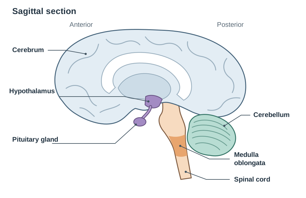
The Brain: Key Regions and Functions
The brain and spinal cord are protected by three membranes called meninges and cushioned by cerebrospinal fluid (CSF), which absorbs mechanical shock and exchanges nutrients and wastes.
- Cerebrum: The largest part of the brain, divided into left and right hemispheres. It controls conscious thought, memory, intelligence, language, emotions, sensory interpretation, and initiates voluntary muscle contractions.
- Cerebellum: Located at the hindbrain beneath the rear of the cerebrum. It coordinates voluntary muscle movement, precision, and balance, making learned movements smooth and automatic.
- Hypothalamus: Located deep in the centre of the brain. It is the primary control centre for homeostasis, regulating internal conditions like temperature and water balance, and it links the nervous system to the endocrine system by regulating the pituitary gland.
- Medulla oblongata: Located at the base of the brainstem connecting to the spinal cord. It regulates vital, involuntary autonomic functions, including breathing rate, heart rate, and reflexes such as swallowing, coughing, and sneezing.
The Spinal Cord: Internal Anatomy and Functions
The spinal cord extends down through the neural canal of the vertebral column. In cross-section, it shows two distinct tissue zones:
- Inner Grey Matter: A butterfly- or H-shaped central core containing unmyelinated structures: nerve cell bodies, dendrites, synapses, and interneurons.
- Outer White Matter: Surrounding tissue composed of bundles of myelinated axons (tracts) running up to and down from the brain. The fatty myelin sheath gives this tissue its glistening white appearance.
- Central canal: A tiny central tube continuous with the brain ventricles, filled with circulating CSF.
- Spinal roots: Thirty-one pairs of spinal nerves attach to the cord via two distinct roots:
- Dorsal Root: Enters the back of the cord, carrying incoming sensory fibres. It features a visible swelling, the dorsal root ganglion, which houses the cell bodies of sensory neurons.
- Ventral Root: Emerges from the front of the cord, carrying outgoing motor fibres whose cell bodies lie inside the spinal cord's grey matter.
Functions of the Spinal Cord
The spinal cord has two primary functions:
- It is the coordinating centre for spinal reflex actions. The sensory neuron, interneuron, and motor neuron all synapse in the grey matter, so an immediate response can be organised without waiting for the brain.
- It is a conduction pathway. The myelinated tracts of white matter carry impulses up to the brain (sensory information) and down from the brain (motor instructions) to the rest of the body.
2. Neurons: Structure, Specialisation, and Types
A neuron is a specialised cell adapted to generate and transmit electrical nerve impulses. A nerve is a bundle of many nerve fibres (axons and dendrons) held together by connective tissue.
General Neuron Anatomy
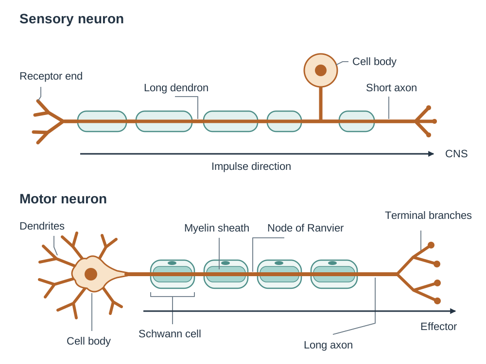
- Cell body: Contains the nucleus, cytoplasm, and dense clusters of mitochondria and ribosomes that synthesise neurotransmitters and supply ATP.
- Dendrites: Short, highly branched cytoplasmic extensions that receive signals from sensory receptors or adjacent neurons and conduct impulses towards the cell body.
- Axon: A single, elongated fibre that carries nerve impulses away from the cell body toward target cells (Axons carry Away).
- Schwann Cells: Specialised cells in the PNS that wrap tightly around the axon during development to produce the myelin sheath.
- Myelin Sheath: A multi-layered, lipid-rich insulating layer produced by Schwann cells. It speeds up the impulse, because it jumps from one gap in the sheath (node of Ranvier) to the next instead of travelling continuously along the whole membrane.
- Axon terminals (synaptic knobs): Bulbous swellings at the distal ends of axon branches containing neurotransmitter vesicles that store chemical messengers.
Impulse Propagation Note: A nerve impulse involves the movement of ions across the neuron membrane. Detailed knowledge of the electrochemistry of ion movement is not required on this syllabus.
Comparing the Three Classes of Neurons
| Characteristic | Sensory Neuron | Interneuron (Relay) | Motor Neuron |
|---|---|---|---|
| Impulse Direction | Receptors CNS | Within CNS (sensory motor) | CNS Effectors (muscles/glands) |
| Cell Body Location | Outside CNS in dorsal root ganglion | Entirely inside CNS grey matter | Inside CNS grey matter (at the beginning of the neuron) |
| Fibre Arrangement | Long receiving dendron, short axon | Short, profusely branched, unmyelinated fibres | Many short dendrites on cell body, long myelinated axon |
Visual Identification on Exam Diagrams
If you are asked to label a diagram, work along the direction of the impulse:
- For a motor neuron: dendrites cell body axon myelin sheath (with Schwann cell) node of Ranvier axon terminals/synaptic knobs with neurotransmitter vesicles. The cell body sits at the start, with short branched dendrites and a long myelinated axon running out to an effector.
- For a sensory neuron: receptor end long dendron cell body on a side branch (in the dorsal root ganglion) short axon leading into the CNS.
3. The Reflex Action and Reflex Arc
A reflex action is a rapid, automatic, involuntary response to a stimulus that does not require conscious brain processing. Reflexes are vital survival adaptations because they protect body tissues from severe injury before the brain has time to register danger or pain.
Named everyday examples of reflex actions include:
- Withdrawing the hand from a hot or sharp object
- The knee-jerk reflex when tapped below the patella
- Blinking when an object moves quickly towards the eyes
- Pupil constriction in response to bright light
- Coughing, sneezing, and swallowing
Not all reflexes are spinal: blinking, pupil constriction, coughing, sneezing and swallowing are coordinated within the brain (swallowing, coughing and sneezing by the medulla oblongata), whereas the withdrawal and knee-jerk reflexes are coordinated locally in the spinal cord.
A reflex arc is the anatomical neural pathway traversed by nerve impulses during a reflex action:
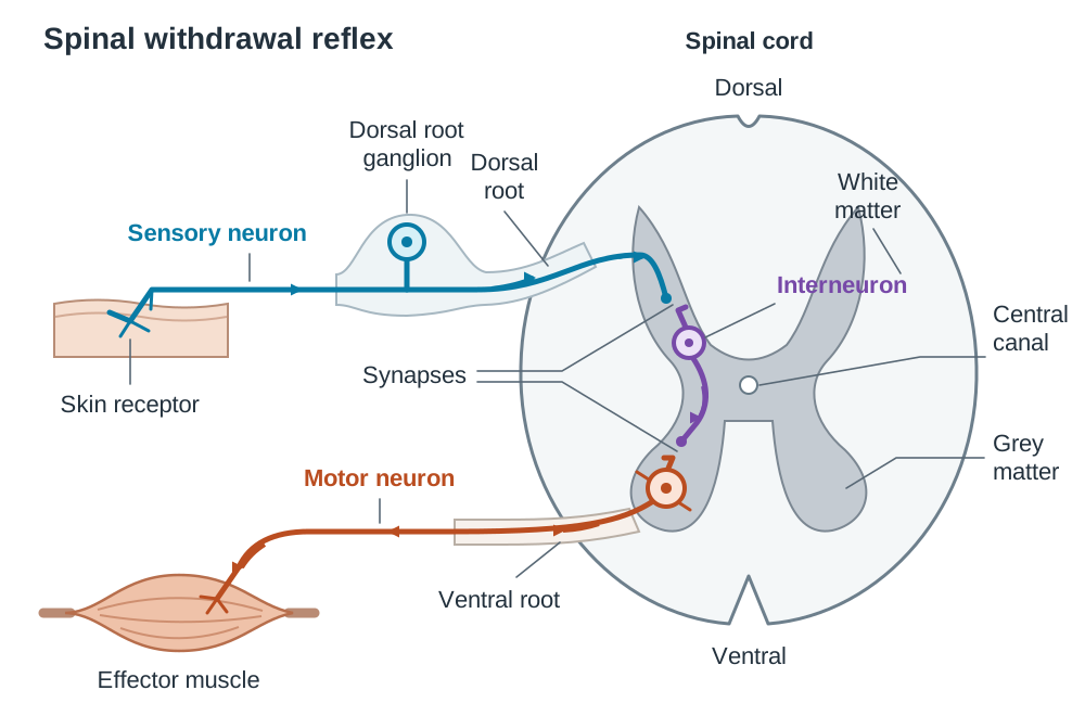
Step-by-Step Pathway of a Spinal Withdrawal Reflex
- Stimulus detection: Intense heat or pressure is detected by pain receptors in the skin, which generate an electrical nerve impulse.
- Sensory transmission: The impulse propagates along the dendron and axon of a sensory neuron, entering the spinal cord via the dorsal root (cell body in the dorsal root ganglion).
- Central integration: Inside the spinal cord's grey matter, the sensory axon releases neurotransmitters across a synapse to stimulate an interneuron.
- Motor output: The interneuron synapses immediately with a motor neuron whose cell body lies in the grey matter. The motor axon emerges from the cord via the ventral root.
- Effector action: The motor axon reaches an effector (such as the biceps muscle), causing the muscle to contract, pulling the limb away from the danger.
Why Pain Perception Follows the Movement
The withdrawal happens first because it is organised locally in the spinal cord across only two synapses. At the same time, collateral impulses travel up the white matter tracts of the spinal cord to the cerebrum. That ascending pathway is longer and involves more synapses, so conscious awareness of pain follows a fraction of a second after the limb has already moved.
4. Homeostasis and Coordination Systems
Homeostasis is the maintenance of a constant internal environment within narrow limits, allowing cells to function properly regardless of external changes. Biological response mechanisms rely on a stimulus-response model that preserves homeostasis via negative feedback.
In a negative feedback loop, any deviation of a physiological condition from its normal set point triggers a corrective response that acts in the opposite direction to restore the balance:
Example 1: Nervous Homeostasis (Thermoregulation)
- Stimulus: Body temperature rises above the set point ().
- Receptors: Thermoreceptors in the skin and internal thermoreceptors in the hypothalamus detect the increase in blood temperature.
- Control centre: The hypothalamus processes the signals and sends nerve impulses along motor neurons to effectors.
- Effectors & response: Cutaneous arterioles dilate (vasodilation) to increase blood flow to the skin surface for heat radiation; sweat glands secrete sweat to cool the skin by evaporation.
- Negative feedback: As blood temperature cools back to normal, hypothalamic stimulation stops, turning off the cooling responses.
Example 2: Hormonal Homeostasis (Blood Glucose Regulation)
- Stimulus: Blood glucose rises above normal after a meal.
- Receptor and control centre: The pancreas detects the rise in blood glucose.
- Endocrine signal: The pancreas secretes the hormone insulin directly into the bloodstream.
- Effectors & response: Liver and muscle cells absorb glucose from the blood and convert it into glycogen for storage.
- Negative feedback: As blood glucose levels fall back to the normal baseline, the pancreas reduces and stops extra insulin secretion.
Comparing Nervous and Hormonal Coordination
Vertebrates maintain homeostasis using both systems side by side:
| Feature | Nervous Coordination | Hormonal (Endocrine) Coordination |
|---|---|---|
| Signalling Form | Electrical impulses along axons and chemical neurotransmitters across synapses | Chemical messengers (hormones) secreted directly into the bloodstream |
| Pathway | Dedicated nerve fibres | Circulated throughout the body in blood plasma |
| Speed of Action | Very fast (milliseconds) | Slower (seconds, hours, or days) |
| Duration of Effect | Short-lived and brief | Long-lasting and sustained |
| Target Area | Highly localised (specific muscle or gland) | Widespread (affects any tissue carrying complementary target receptors) |
| Shared Role | Both link receptors to effectors via a control centre, and both rely on negative feedback |
5. Synaptic Transmission
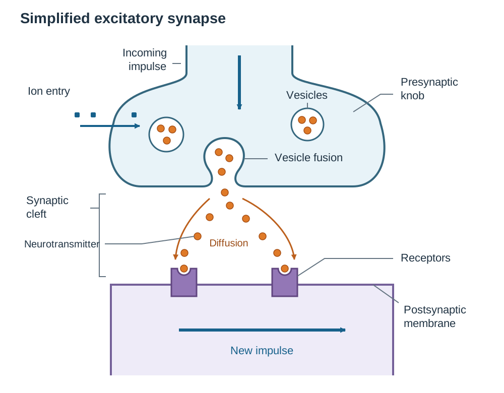
A synapse is the functional junction where two neurons come into close proximity to pass on information. The fluid-filled microscopic space separating them is the synaptic cleft. Because an electrical impulse cannot jump across this fluid gap, communication is converted into a chemical signal and back into an electrical signal.
Step-by-Step Mechanism of Synaptic Transmission
- An electrical nerve impulse arrives at the synaptic knob of the presynaptic neuron.
- The impulse causes an influx of ions across the presynaptic membrane.
- The ions cause the neurotransmitter vesicles to move to the presynaptic membrane and fuse with it, releasing neurotransmitter into the cleft by exocytosis, where it stays only for a short time.
- Neurotransmitter molecules diffuse across the fluid-filled synaptic cleft down their concentration gradient.
- The neurotransmitter binds to specific complementary protein receptors on the postsynaptic membrane.
- This binding causes ion channels to open, which starts a new electrical nerve impulse in the next (postsynaptic) neuron.
- The neurotransmitter is then rapidly cleared from the cleft to prevent continuous stimulation of the postsynaptic neuron:
- Enzymatic Inactivation: An enzyme in the cleft breaks the neurotransmitter down into inactive fragments. For example, the enzyme acetylcholinesterase breaks down acetylcholine. The fragments are reabsorbed by the presynaptic knob to resynthesise fresh neurotransmitter.
- Reuptake: Intact neurotransmitter molecules (such as dopamine or serotonin) are transported directly back into the presynaptic neuron by active transport pumps to be repackaged into vesicles.
Significance of Synapses
- Directional control: Synapses ensure impulses travel in one direction only. Neurotransmitter vesicles exist exclusively in presynaptic knobs, and complementary receptors are found exclusively on postsynaptic membranes.
- Prevention of overstimulation: If a neuron is continuously stimulated, its store of neurotransmitter vesicles can become depleted faster than they are reformed, temporarily halting transmission and preventing fatigue in effectors.
6. Synaptic Disruptions, Pharmacology, and Lifestyle (Higher Level)
Proper nervous coordination requires the exact regulation of impulse transmission and neurotransmitter turnover. Disruptions can occur physically along the nerve fibre or chemically at the synapse.
Physical Disruptions Along Nerve Fibres
- Multiple Sclerosis (MS): An autoimmune disorder in which the body's immune system attacks and destroys the myelin sheath around axons in the CNS. Without this insulating fatty layer, electrical impulses leak across the membrane and can no longer jump between the nodes of Ranvier. Conduction slows down drastically or is blocked entirely, resulting in muscle weakness, poor coordination, numbness, and blurred vision. Treatments focus on immunosuppressive medications to reduce attacks and physiotherapy; lost myelin cannot currently be restored.
- Paralysis: Loss of voluntary muscle movement caused by nerve impulses failing to reach effector muscles. A common cause is physical trauma to the vertebral column that severs descending motor tracts in the spinal cord's white matter. Motor impulses from the brain cannot travel past the site of damage, so voluntary movement below the injury is lost. Treatment involves spinal stabilisation surgery, assistive devices, and physiotherapy. Note that spinal reflexes below the injury may remain active because the local spinal reflex arc is intact.
Pharmacological Disruptions at the Synapse
- Enzyme Inhibitors: Certain chemicals block the enzymes responsible for clearing neurotransmitters. Organophosphate insecticides and nerve agents inhibit acetylcholinesterase, causing acetylcholine to accumulate in the synaptic cleft. This leads to continuous, uncontrolled stimulation of muscle receptors, causing spasms and respiratory paralysis.
- Reuptake Inhibitors: Selective Serotonin Reuptake Inhibitors (SSRIs) block the transport proteins that pump serotonin back into the presynaptic neuron. Serotonin remains in the cleft for longer, enhancing postsynaptic firing to treat clinical depression.
- Painkillers (Analgesics): Medications that interfere with pain pathways across synapses. Some bind to postsynaptic receptors without triggering an impulse, blocking neurotransmitters from binding. Opioids (such as morphine) bind to presynaptic receptors, inhibiting the release of pain neurotransmitters.
Roles of Dopamine and Endorphins
- Dopamine: Operates in the brain's reward pathway, motivation, emotional regulation, and voluntary motor coordination. Parkinson's disease is caused by the gradual death of dopamine-producing neurons in the brain (in a region called the substantia nigra), which reduces the supply of dopamine to the brain areas that coordinate movement. This produces muscle tremors, stiffness, and slowness of movement. It is treated using L-dopa (which crosses the blood-brain barrier and is converted to dopamine) or drugs that inhibit dopamine-degrading enzymes.
- Endorphins: Neuropeptides produced by the pituitary gland and hypothalamus that act as natural painkillers. They inhibit pain perception in the brain and spinal cord and induce feelings of calm and wellbeing.
Influence of Lifestyle Choices
- Physical exercise: Sustained aerobic exercise stimulates the release of both endorphins and dopamine. This suppresses pain, lifts mood naturally, and reduces stress.
- Dietary choices: Dopamine is made in the body from the amino acid tyrosine, which the body can produce from the essential amino acid phenylalanine obtained in the diet. A diet with enough protein (lean meat, fish, eggs, dairy, legumes) supplies the raw material for dopamine synthesis.
- Non-medical drug use: Recreational drugs such as cocaine, nicotine, and opioids flood synapses with dopamine or mimic endorphins. Prolonged drug use leads to cellular adaptations: the postsynaptic membrane downregulates (decreases) its receptor count and natural transmitter synthesis drops. The user develops tolerance (needing higher doses to get the same effect) and physical dependence, experiencing withdrawal symptoms when the drug is removed.
Link to the Biology in Practice Investigation
This topic links naturally to the unifying strand and practical investigations on stimulus and response. For example, testing human reaction time using a dropped-ruler experiment generates reliable primary data. A testable hypothesis might state: Reaction time to a visual stimulus decreases with repeated practice over ten trials. When designing this experiment, consider key quality descriptors: fairness (identical ruler, consistent starting finger gap, same subject), repeatability (multiple repeats and calculating a mean), identifying sources of error (anticipating the drop), and explaining any anomalous data points.
Key terms
- Stimulus
- Any change in the internal or external environment that produces a response in an organism.
- Receptor
- A cell, tissue, or organ that detects a stimulus and converts it into an electrical nerve impulse.
- Effector
- A muscle or gland that carries out a bodily response to a stimulus.
- Nerve Impulse
- An electrical signal that travels along a neuron, involving the movement of ions across the neuron membrane.
- Central Nervous System (CNS)
- The brain and spinal cord, serving as the body's central processing and command centre.
- Peripheral Nervous System (PNS)
- All nervous tissue outside the central nervous system, consisting of nerve fibres and ganglia.
- Neuron
- A specialised cell adapted to generate and transmit electrical nerve impulses.
- Ganglion
- A swelling located outside the central nervous system containing a cluster of nerve cell bodies.
- Myelin Sheath
- A lipid-rich insulating layer produced by Schwann cells around an axon that accelerates nerve impulse transmission.
- Schwann Cell
- A specialised cell in the peripheral nervous system that wraps around an axon to produce the myelin sheath.
- Reflex Action
- A rapid, automatic, involuntary response to a stimulus that does not require conscious intervention.
- Reflex Arc
- The anatomical pathway traversed by nerve impulses during a reflex action (receptor, sensory neuron, interneuron, motor neuron, effector).
- Homeostasis
- The maintenance of a constant internal environment within narrow physiological limits.
- Negative Feedback
- A self-regulating control mechanism where a detected deviation triggers a response that directly opposes and reverses the change to restore equilibrium.
- Synapse
- The functional region where two neurons (or a neuron and an effector) come into close proximity to transfer information.
- Synaptic Cleft
- The fluid-filled microscopic space separating the presynaptic knob from the postsynaptic membrane.
- Neurotransmitter
- A chemical messenger stored in synaptic vesicles that diffuses across the synaptic cleft to transmit an impulse to the next cell.
- Reuptake
- The active transport of intact neurotransmitter molecules from the synaptic cleft back into the presynaptic neuron.
- Dopamine
- A neurotransmitter that functions in the brain's reward pathway, motivation, emotional regulation, and voluntary motor control.
- Endorphins
- Neuropeptides produced in the brain and pituitary gland that act as natural painkillers to suppress pain and enhance wellbeing.
Check yourself
Which specific brain structure connects the nervous and endocrine systems and acts as the master control centre for homeostasis?
The hypothalamus.
State the two primary functions of the spinal cord.
It serves as the coordinating centre for spinal reflex actions (in grey matter) and acts as a two-way conduction pathway carrying impulses to and from the brain (in white matter).
Where are the cell bodies of sensory neurons and motor neurons located relative to the spinal cord?
Sensory neuron cell bodies are located outside the CNS in the dorsal root ganglion; motor neuron cell bodies are located inside the CNS within the spinal cord's grey matter.
List two named examples of reflex actions coordinated by the brain and two coordinated by the spinal cord.
Brain reflexes: blinking and pupil constriction (or coughing/swallowing, coordinated by the medulla). Spinal reflexes: withdrawal reflex and knee-jerk reflex.
Why can nerve impulses only travel in one direction across a chemical synapse?
Because neurotransmitter vesicles are present only in the presynaptic terminal knob, and complementary neurotransmitter receptors are present only on the postsynaptic membrane.
A person's myelin sheaths are being damaged by their own immune system in multiple sclerosis. Explain why their muscle responses become slow or uncoordinated.
Myelin insulates the axon and allows impulses to jump between nodes of Ranvier; when it is destroyed, impulses leak across the membrane and travel much more slowly or are blocked entirely, delaying motor signals to the effector muscles.
How does an acetylcholinesterase inhibitor drug affect synaptic transmission at a neuromuscular junction?
It blocks the breakdown of acetylcholine in the cleft, causing it to remain longer and at higher concentrations, which results in continuous or enhanced stimulation of postsynaptic receptors.
From which amino acid is dopamine synthesised in the body, and what dietary requirement is needed for its production?
Dopamine is made from tyrosine, which the body synthesises from the essential dietary amino acid phenylalanine; therefore, adequate protein in the diet is required.