

The human body responds to environmental changes using four coordinated systems: the nervous system, the endocrine system, the immune system, and the musculoskeletal system. In this response pathway, skeletal muscles act as effectors at the end of a reflex or voluntary chain, contracting to pull on bones across movable joints. This topic covers the structure and functions of the axial and appendicular skeletons, the roles of cartilage, ligaments, and tendons in synovial joints, the action of antagonistic muscle pairs, and how to construct, test, and evaluate a physical model of limb movement.
Response Systems and the Functions of the Skeleton
The musculoskeletal system provides the physical machinery for bodily response. When a receptor detects an internal or external stimulus, nerve impulses travel along a sensory neuron into the central nervous system, which directs impulses out along a motor neuron to an effector. Skeletal muscles are effectors. When stimulated, they contract and pull on bones across joints to produce movement.
For example, if you step on a sharp stone: the stimulus (sharp pressure) stimulates pain receptors in the skin of the foot, sending impulses along a sensory neuron to the spinal cord. An interneuron passes the impulse to a motor neuron, which stimulates leg muscles (the effectors) to contract and pull the foot away. Bones, muscles, tendons, and joints act as a unified response system.
Four Functions of the Skeleton
- Support — forms a rigid internal framework that keeps the body upright and maintains its shape.
- Protection — hard bone encloses soft vital organs: the cranium shields the brain, the vertebral column protects the spinal cord, the ribs and sternum protect the heart and lungs, and the pelvic girdle protects the lower abdominal and reproductive organs.
- Movement — bones act as rigid levers that skeletal muscles pull across joints.
- Blood cell production — red bone marrow, found inside bones such as the sternum, ribs, and vertebrae, produces red blood cells, white blood cells, and platelets.
Bone also acts as the body's main reservoir for essential minerals, storing calcium and phosphorus that can be mobilised into the bloodstream when required.
The Axial Skeleton: Skull, Vertebrae, Ribs, and Sternum
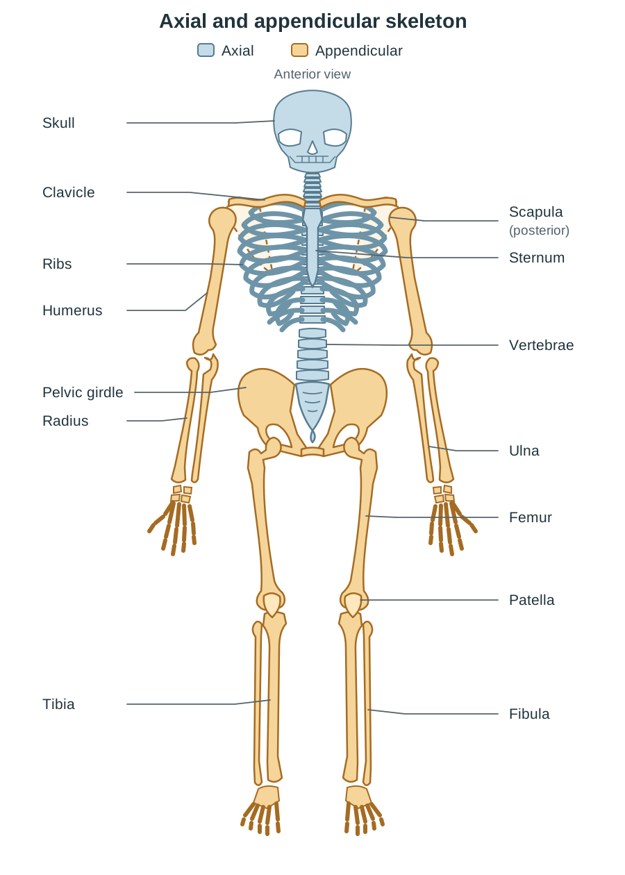
The axial skeleton forms the central longitudinal axis of the human body. It consists of four main groups of bones: the skull, vertebrae, ribs, and sternum. Between the vertebrae lie the intervertebral discs, which are specialized pads of fibrocartilage rather than bones.
| Skeletal component | Structure and position | How structure relates to function |
|---|---|---|
| Skull | Composed of the fused bones of the cranium and facial bones joined at immovable joints called sutures. | The hard, rounded cranium encloses and shields the brain from mechanical trauma. Facial bones form rigid protective sockets for sensory organs such as the eyes. |
| Vertebrae | A series of bones stacked vertically to form the vertebral column. Each vertebra has a thick anterior body, a central vertebral canal, and bony processes. | The solid vertebral body bears body weight; the aligned openings form a continuous bony canal that encloses and protects the delicate spinal cord; bony processes provide anchor points for muscles and ligaments. |
| Intervertebral discs | Discs of tough fibrocartilage positioned between adjacent vertebral bodies. | They act as shock absorbers, cushioning compressive loads during walking, running, and jumping. They allow slight movement between neighbouring vertebrae, giving the spine overall flexibility while safeguarding the spinal cord. |
| Ribs | Twelve pairs of curved bones. Each pair joins the thoracic vertebrae at the back and curves around the chest toward the front. | Their curved structure forms a protective cage around the heart and lungs. By pivoting upward and downward, they change the volume of the thoracic cavity during breathing. |
| Sternum | A flat dagger-shaped bone running down the anterior midline of the chest. | It anchors the ribs firmly at the front of the chest through costal cartilages, protects the heart and lungs behind it, and contains red bone marrow that makes blood cells. |
The Appendicular Skeleton: Girdles and Limbs
The appendicular skeleton consists of the pectoral and pelvic girdles together with the attached bones of the upper and lower limbs. Its primary adaptations are for locomotion, posture, and manual dexterity.
The Girdles: Mobility versus Stability
A girdle anchors a limb system to the central axial skeleton:
- Pectoral girdle: Consists of the clavicle (collarbone) and scapula (shoulder blade) on each side. The clavicle is a slender strut holding the shoulder away from the chest. The scapula is a flat triangular bone providing a broad surface for muscle attachment and a shallow socket for the arm. The pectoral girdle's only bony attachment to the axial skeleton is where the clavicle meets the sternum; the scapula itself is held in place mainly by muscles. This loose arrangement permits a wide range of arm movement, though it makes the shoulder joint more prone to dislocation.
- Pelvic girdle: A heavy, rigid ring of bone fused directly to the sacrum of the vertebral column. It supports upper body weight and transmits locomotion forces into the legs. Its deep cup-like socket provides immense joint stability for the hip. Note: Under the Leaving Certificate specification, you do not need to name the individual fused bones that make up the pelvic girdle.
The Attached Limbs
The upper and lower limbs are built to the same basic architectural plan — one long proximal bone, followed by two distal bones — but each is adapted for a distinct purpose:
| Region | Upper limb | Lower limb | Structural and functional comparison |
|---|---|---|---|
| Upper (proximal) bone | Humerus | Femur | The humerus is a mobile arm lever articulating with the shallow scapular socket; the femur is the longest, strongest bone in the body, designed to support full body weight within the deep pelvic socket. |
| Lower (distal) bones | Radius & Ulna | Tibia & Fibula | In the forearm, the radius lies on the thumb side and rotates over the ulna to turn the hand, while the ulna forms the elbow hinge. In the lower leg, the thick tibia bears weight down to the ankle, while the slender fibula provides attachment points for muscles and stabilises the ankle. |
- Patella (kneecap): A specialized bone embedded within the tendon of the thigh muscles at the front of the knee. It protects the knee joint and improves the pulling leverage of the quadriceps muscle when straightening the leg. The upper limb has no equivalent bone.
Synovial Joints and Connective Tissues
A joint occurs where two or more bones meet. Joints are classified by mobility into immovable joints (such as skull sutures), slightly movable joints (such as cartilaginous joints between vertebrae), and freely movable synovial joints (such as the elbow, knee, shoulder, and hip).
Types of Synovial Joints
- Hinge joint (e.g. the elbow and knee): Allows movement in a single plane, specifically flexion (bending) and extension (straightening).
- Ball-and-socket joint (e.g. the shoulder and hip): The rounded head of one bone fits into a cup-shaped socket of another, allowing movement in all planes as well as rotation.
Diagram Checklist: A Synovial Joint
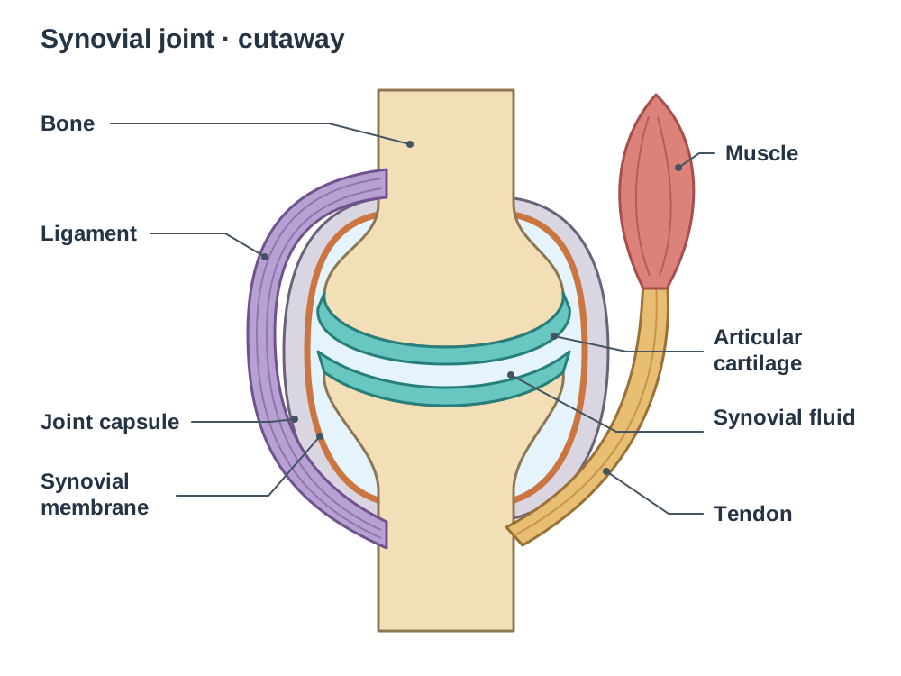
When sketching and labelling a synovial joint (such as the knee or elbow), include these seven structures with their functions:
- Bone — provides rigid structural levers for movement.
- Articular cartilage — smooth layer capping bone ends; absorbs shock and reduces friction.
- Joint capsule — tough fibrous sleeve enclosing the joint; holds fluid in and protects internal structures.
- Synovial membrane — thin inner connective tissue lining of the capsule; secretes synovial fluid.
- Synovial fluid — lubricating liquid filling the cavity; reduces friction and nourishes living cartilage cells.
- Ligament — dense fibrous band running bone to bone; holds bones aligned and prevents dislocation.
- Tendon — dense fibrous band running muscle to bone; transfers muscle pulling force directly to bone.
Connective Tissue Comparison
| Connective tissue | Connection | Physical property | Primary function |
|---|---|---|---|
| Cartilage | Covers articulating bone ends; forms intervertebral discs | Smooth, firm, collagen matrix; avascular (lacks blood vessels) | Absorbs compressive shock and reduces friction between moving bone ends. |
| Ligament | Bone to bone | Tough fibrous bands; slightly elastic | Holds articulating bones in place, stabilises the joint, and prevents abnormal movements or dislocation. |
| Tendon | Muscle to bone | Dense cords of parallel collagen fibres; inelastic | Transmits the pulling tension of muscle contraction directly to bone without stretching. |
Because cartilage is avascular, oxygen and nutrients reach cartilage cells exclusively by slow diffusion from synovial fluid. Consequently, worn or torn cartilage (such as in osteoarthritis) repairs far more slowly than vascularised bone.
Antagonistic Muscle Pairs: Mechanics of Movement
Skeletal muscles produce movement through muscular contraction, an active process powered by ATP. When a muscle contracts, its fibres shorten, generating pulling tension that is transferred to bone via tendons. Crucially, muscles can only pull — they can never push.
Because a contracted muscle cannot push a bone back into its resting position, joints require an antagonistic muscle pair: two muscles that work in opposition across the same joint. When one muscle contracts, the other relaxes and is passively stretched.
Attachment of a Muscle: Origin and Insertion
A skeletal muscle is attached to bone by a tendon at each end:
- The origin is attached to the bone that remains relatively stationary during contraction.
- The insertion is attached to the bone that moves.
When a muscle contracts and shortens, it pulls its insertion toward its origin.
The Biceps and Triceps Mechanism (Elbow Joint)
The biceps brachii and triceps brachii form the standard antagonistic pair operating the elbow hinge:
- Flexion (bending the elbow):
The biceps brachii acts as the flexor (it decreases the angle at the joint). The biceps has its origin on the scapula and humerus, and its insertion on the radius. When the biceps contracts and shortens, it pulls on the radius. Simultaneously, the triceps brachii (the extensor) relaxes and is passively stretched. The forearm rises toward the upper arm.
- Extension (straightening the elbow):
The triceps brachii acts as the extensor (it increases the angle at the joint). The triceps has its origin on the humerus and scapula, and its insertion on the ulna at the point of the elbow. When the triceps contracts and shortens, it pulls on the ulna. Simultaneously, the biceps brachii relaxes and is stretched. The forearm lowers until the arm is straight.
[Flexion]   Biceps CONTRACTS (pulls radius) + Triceps RELAXES  -> Forearm raises
[Extension] Triceps CONTRACTS (pulls ulna)   + Biceps RELAXES   -> Forearm lowers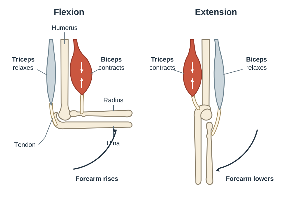
The same antagonistic principle operates at the knee: the quadriceps on the front of the thigh contracts to straighten the leg while the hamstrings relax; the hamstrings on the back of the thigh contract to bend the knee while the quadriceps relaxes.
Modelling an Antagonistic Muscle Pair (Higher Level)
At Higher Level, learning outcome 2.4.3 requires you to model the function of an antagonistic muscle pair. A scientific model is a simplified representation of a biological system used to explain mechanisms, demonstrate principles, and make testable predictions.
A classic mechanical arm model uses two rigid battens hinged by a bolt, controlled by two opposing pull-cords:
| Model component | Biological analogue | Biological role represented |
|---|---|---|
| Fixed upper batten | Humerus (upper arm) | Stationary bone providing the site of muscle origin. |
| Movable lower batten | Radius and ulna (forearm) | Lever arm carrying the insertion point, rotating at the joint. |
| Pin / bolt hinge | Elbow joint | Pivot fulcrum allowing rotation in a single plane. |
| Front pull-cord | Biceps muscle and tendon | Flexor generating pulling tension to raise the forearm. |
| Back pull-cord | Triceps muscle and tendon | Extensor generating pulling tension to straighten the forearm. |
[Model Summary]
Pull front cord = flexor contracts -> lower strip rises;
Pull back cord  = extensor contracts -> lower strip straightens;
Pin = hinge joint;
External hand supplies the energy; ATP supplies it in the body.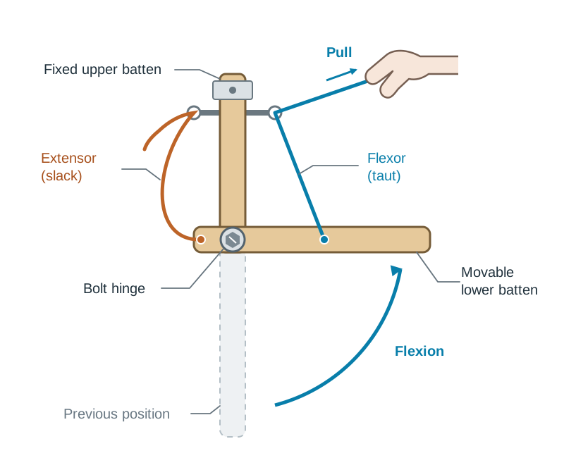
Making and Testing Predictions
- Testable prediction: If the front pull-cord is cut or detached from the lower batten, pulling on it produces no upward movement. This accurately predicts that tearing a biceps tendon prevents active flexion of the arm, even if the muscle belly contracts.
Biological Limitations of the Model
Every model oversimplifies biological reality:
- Active contraction versus manual tension: In the model, tension is generated externally by pulling a cord. In living muscle, contraction occurs from within muscle fibres consuming cellular ATP in response to motor nerve impulses.
- Absence of lubricating tissues: The metal bolt lacks articular cartilage, a synovial membrane, and synovial fluid, failing to illustrate biological lubrication and shock absorption.
- Absence of stabilising ligaments: The model relies on a rigid bolt rather than flexible ligaments connecting bone to bone, so it cannot demonstrate joint stability or dislocation.
Key terms
- Axial Skeleton
- The central core of the skeleton, consisting of the skull, vertebrae, ribs, and sternum.
- Appendicular Skeleton
- The bones of the upper and lower limbs together with the pectoral and pelvic girdles that attach them to the axial skeleton.
- Intervertebral Disc
- A pad of fibrocartilage located between adjacent vertebrae that cushions mechanical shock and permits slight spinal flexibility.
- Pectoral Girdle
- The skeletal structure formed by the clavicle and scapula that attaches the upper limb to the axial skeleton.
- Pelvic Girdle
- A heavy ring of bone fused to the sacrum that attaches the lower limbs to the axial skeleton and supports body weight.
- Synovial Joint
- A freely movable joint enclosed by a fibrous capsule lined with a synovial membrane that secretes lubricating synovial fluid.
- Articular Cartilage
- A smooth, firm layer of connective tissue covering bone ends in synovial joints that reduces friction and absorbs shock.
- Ligament
- A tough, slightly elastic band of fibrous connective tissue that connects bone to bone across a joint to provide stability.
- Tendon
- A tough, inelastic cord of fibrous connective tissue that connects muscle to bone, transmitting the pulling force of contraction.
- Antagonistic Muscle Pair
- A pair of muscles that produce opposite physical movements across the same joint; when one contracts, the other relaxes.
- Flexor
- A muscle that contracts to decrease the angle between articulating bones at a joint (such as the biceps bending the elbow).
- Extensor
- A muscle that contracts to increase the angle between articulating bones at a joint (such as the triceps straightening the elbow).
- Origin
- The end of a skeletal muscle attached to the stationary bone during contraction.
- Insertion
- The end of a skeletal muscle attached to the movable bone that is pulled during contraction.
Check yourself
Name the bones of the axial skeleton, and state what structure lies between the vertebrae.
The bones are the skull, vertebrae, ribs, and sternum; pads of fibrocartilage called intervertebral discs lie between the vertebrae.
State four general functions of the human skeleton.
Support, protection of vital organs, movement, and blood cell production (it also serves as a mineral store for calcium and phosphorus).
Why is the pelvic girdle attached more firmly to the axial skeleton than the pectoral girdle?
The pelvic girdle must bear the weight of the upper body and transfer large forces to the legs for walking and running, requiring stability rather than high mobility.
Distinguish between a ligament and a tendon by stating the structures each connects.
A ligament connects bone to bone, whereas a tendon connects muscle to bone.
Why does damaged articular cartilage repair much more slowly than fractured bone?
Cartilage is avascular (lacks blood vessels), so oxygen and nutrients must reach the cells slowly by diffusion from surrounding tissues and synovial fluid.
What happens to the biceps and triceps when you actively straighten your elbow?
The triceps (extensor) contracts and shortens, pulling on the ulna, while the biceps (flexor) relaxes and is stretched.
Give one way a mechanical pull-cord model misrepresents how living muscle generates force.
The model uses an external manual pull on passive strings, whereas living muscle generates pulling force internally using ATP in response to motor nerve impulses.
Give one biological structure of a real synovial joint that a simple pin hinge cannot represent.
The pin hinge lacks articular cartilage, a synovial membrane, synovial fluid, and stabilising ligaments.