

DNA replication and protein synthesis represent the core mechanisms of molecular genetics. During interphase of the cell cycle, DNA replicates semiconservatively so that each dividing cell receives an identical and complete set of genetic instructions. Gene expression converts this stored code into functional proteins through a two-step pathway: transcription of a DNA template strand into messenger RNA (mRNA) in the nucleus, followed by translation of the mRNA codons into a specific sequence of amino acids at the ribosome. Changes in the DNA base sequence or chromosome structure produce mutations. These can alter protein function or disrupt normal cell-cycle control, leading to uncontrolled mitotic division and cancer.
The Genetic Code, Genes, and Traits
A gene is a section of DNA that codes for the production of a specific protein or polypeptide. Genetic instructions dictate an organism's characteristics through a clear chain of cause and effect:
- The sequence of bases along a gene determines the sequence of codons in mRNA.
- The codon sequence determines the sequence of amino acids joined at the ribosome.
- The amino acid sequence dictates how the polypeptide folds into its three-dimensional shape, determining protein function.
- The functional protein produces the physical or physiological trait (characteristic).
For example, the gene for the enzyme tyrosinase codes for the production of functional tyrosinase. This enzyme converts tyrosine into melanin, the pigment responsible for skin, hair, and eye colour. A mutation in this gene can produce an inactive enzyme; without melanin, the organism displays the trait of albinism. This pathway is summarised as: DNA mRNA protein trait.
The Triplet Code and tRNA Structure
Living organisms use 20 different amino acids to construct proteins, but DNA contains only 4 distinct nitrogenous bases (adenine, thymine, guanine, and cytosine). A single-base code could only specify 4 amino acids (). A two-base doublet code could specify only 16 (), which is still insufficient. Cells therefore rely on a triplet code ( combinations), which provides more than enough codons to code for all 20 amino acids as well as start and stop instructions. Because several codons can code for the same amino acid, the code is described as degenerate.
In mRNA, each triplet is called a codon. Transfer RNA (tRNA) acts as the physical adaptor between the codon sequence and the amino acid sequence. Each tRNA molecule is folded into a cloverleaf shape held by hydrogen bonds. At one end, it carries an exposed three-base sequence called an anticodon, which matches a specific mRNA codon by complementary base pairing. At the opposite end, it carries an attachment site for the specific amino acid corresponding to that codon. Because each anticodon is exactly three bases long, one tRNA molecule brings precisely one amino acid for each codon read by the ribosome.
Nucleic Acid Chemistry
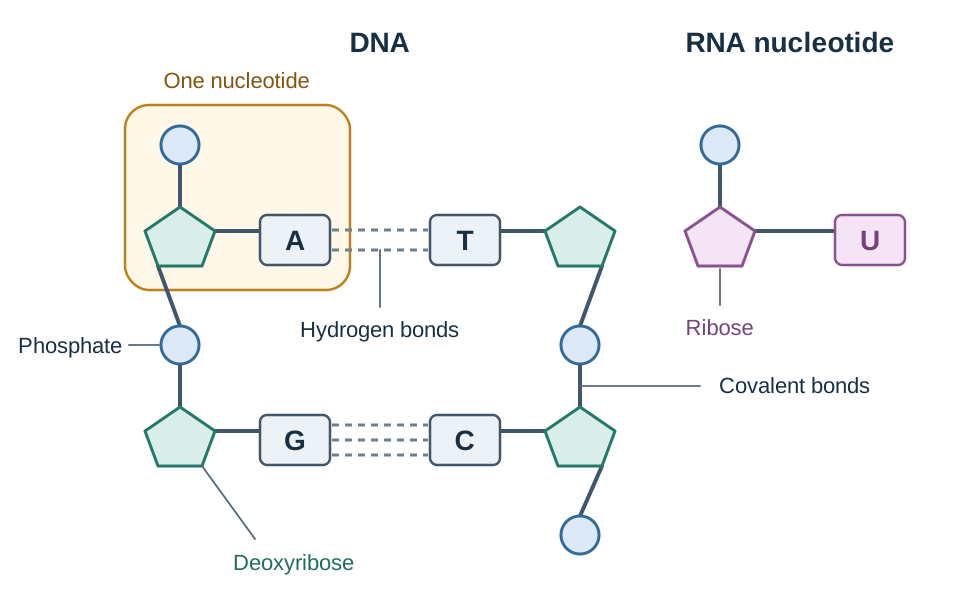
DNA and RNA are polymers made of repeating monomers called nucleotides. Each nucleotide contains:
- A five-carbon pentose sugar (deoxyribose in DNA, ribose in RNA)
- A phosphate group
- A nitrogenous base
The bases are divided into double-ring purines (adenine and guanine) and single-ring pyrimidines (thymine and cytosine in DNA; uracil and cytosine in RNA). In DNA, adenine pairs with thymine via two hydrogen bonds (), while guanine pairs with cytosine via three hydrogen bonds (). In RNA, adenine pairs with uracil ().
Semiconservative DNA Replication and the Cell Cycle
DNA replication occurs during interphase of the cell cycle, before nuclear division begins. Replication is essential so that each chromosome is duplicated into two identical sister chromatids joined at the centromere. During mitosis, these chromatids are separated into opposite daughter cells. Without prior replication, each new cell would receive only half the necessary genetic instructions. Furthermore, any copying mistakes made during replication become permanent and are passed to all descendant cells.
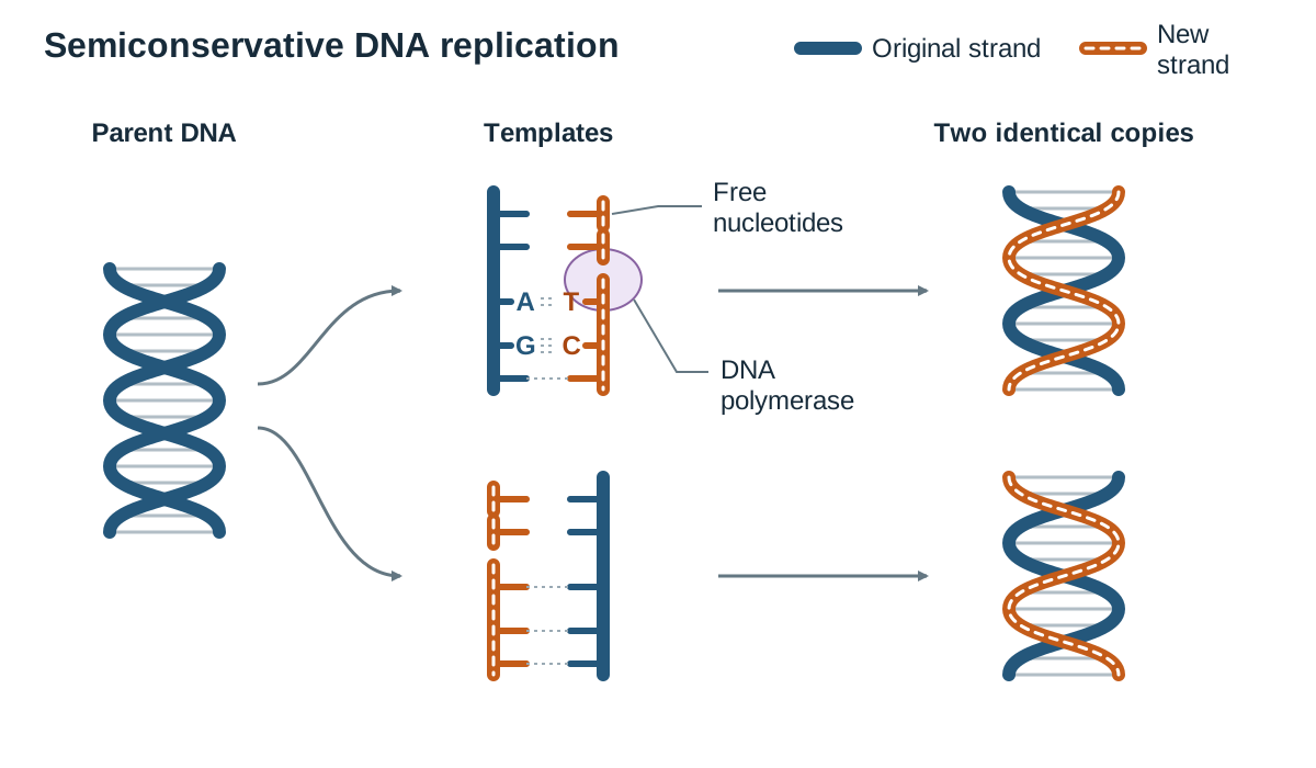
Replication follows a semiconservative mechanism because each newly formed double helix conserves one original parent strand and incorporates one newly made daughter strand.
Steps in DNA Replication
- Enzymes break the hydrogen bonds between base pairs, unzipping the parent double helix and exposing the bases on both template strands.
- Free DNA nucleotides present in the nucleoplasm move to the exposed template strands and align opposite their complementary bases ( with , and with ).
- The enzyme DNA polymerase joins adjacent nucleotides by building the covalent sugar-phosphate backbone of the new strand. DNA polymerase also proofreads the newly added bases, correcting mismatched pairs.
- Each new double-stranded molecule rewinds into a double helix, producing two genetically identical copies of the original DNA.
Scientific Models of DNA Replication
Biologists use diagrams and physical models to represent replication. A typical diagram shows the parent double helix partially unzipped into a 'Y-shaped' replication fork, with free nucleotides aligning along each template strand and DNA polymerase moving behind them, resulting in two shaded strands (one old, one new). While useful, these models have limitations:
- They are drawn flat like a ladder, ignoring the natural three-dimensional twist of the double helix.
- They show only a handful of bases, whereas real human chromosomes contain millions of base pairs.
- They depict replication as a static, step-by-step event, whereas in living cells it is a continuous, rapid biochemical process involving many coordinating enzymes.
Transcription: Synthesising Messenger RNA
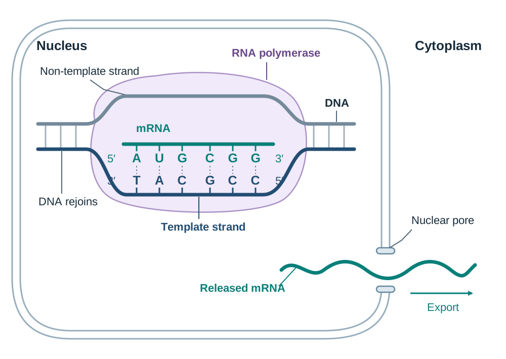
Transcription is the first stage of protein synthesis. It takes place in the nucleus, where the genetic code of a specific gene on DNA is transcribed into a single strand of messenger RNA (mRNA).
Mechanism of Transcription
- The enzyme RNA polymerase binds to the DNA at the start of a gene (a specific base sequence called a promoter).
- RNA polymerase unwinds the double helix and breaks the hydrogen bonds between complementary bases, exposing the template strand.
- Free RNA nucleotides present in the nucleoplasm align opposite their complementary bases on the DNA template strand: cytosine pairs with guanine, guanine pairs with cytosine, thymine on DNA pairs with adenine on RNA, and adenine on DNA pairs with uracil on RNA.
- RNA polymerase joins the RNA nucleotides together to form the continuous single strand of mRNA.
- When RNA polymerase reaches a termination signal at the end of the gene, the newly formed mRNA transcript detaches.
- The DNA template strand re-bonds with its partner strand and rewinds into a double helix. The mRNA transcript leaves the nucleus through a nuclear pore and enters the cytoplasm.
Translation: Assembling the Polypeptide Chain
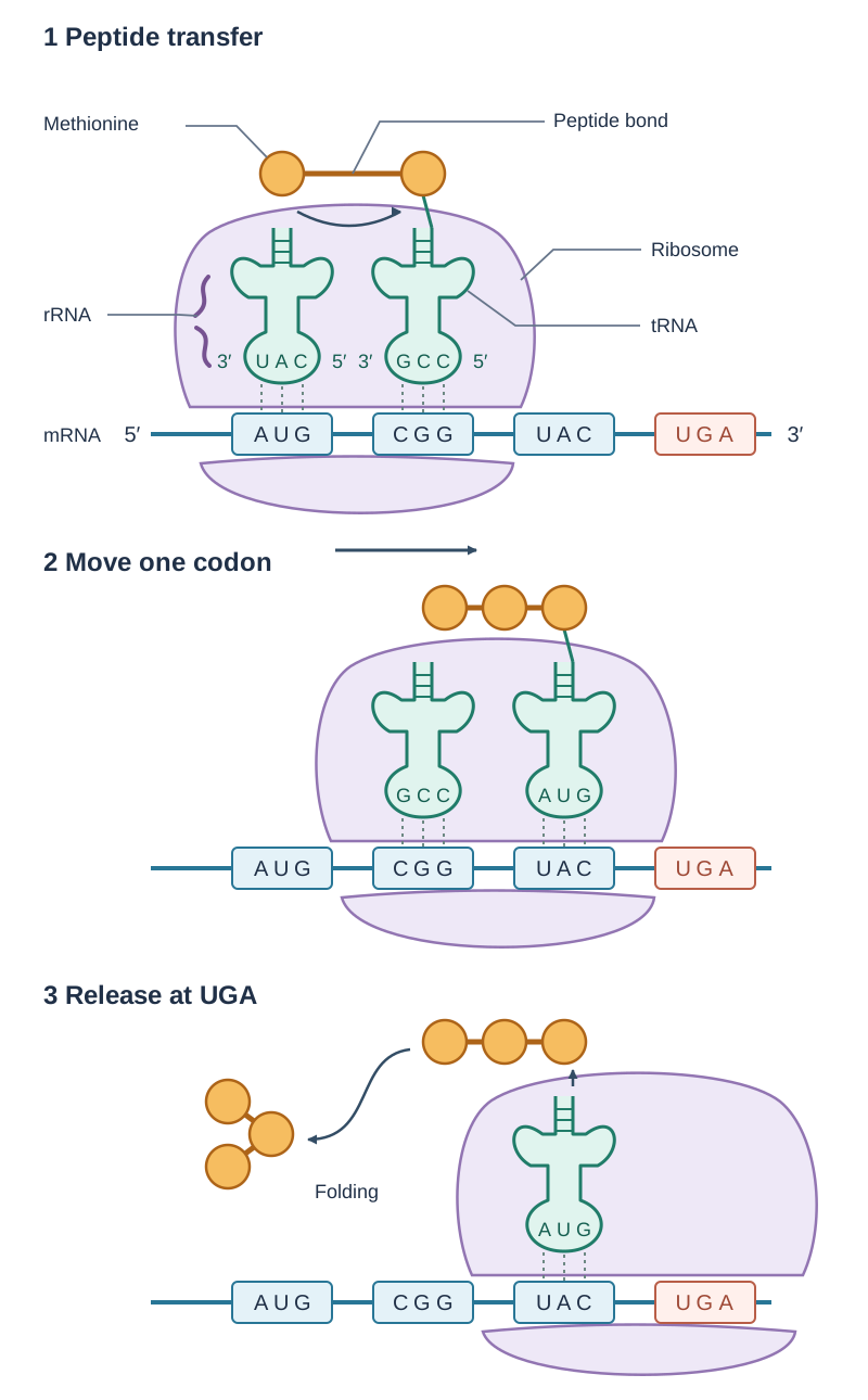
Translation converts the codon sequence of an mRNA molecule into a specific sequence of amino acids joined by peptide bonds. This process takes place at ribosomes in the cytoplasm.
Steps in Translation
- Initiation: The mRNA strand docks onto a ribosome. The ribosome reads the strand until it locates the start codon (AUG). An initiator tRNA carrying the amino acid methionine binds to the start codon using its complementary anticodon (UAC).
- Elongation: The ribosome moves along the mRNA, reading one codon at a time. A second tRNA carrying its matching amino acid binds to the adjacent codon. The ribosome (its rRNA) catalyses the formation of a peptide bond between neighbouring amino acids. Once the bond forms, the first tRNA releases its amino acid and leaves the ribosome to collect another free amino acid in the cytoplasm. The ribosome then steps forward by one codon.
- Termination and Folding: Elongation continues until the ribosome reaches one of the three stop codons: UAA, UAG, or UGA. A stop codon does not code for an amino acid, and no tRNA molecule possesses a matching anticodon. Translation ceases, the completed polypeptide chain detaches from the ribosome, and it folds into its functional three-dimensional shape (such as a fibrous structural protein or a globular enzyme).
Modelling Translation
A standard model of translation shows the mRNA passing between ribosomal subunits, labelled codons grouped in threes, tRNAs docking at adjacent sites, and a growing chain of amino acid circles connected by peptide bonds. Like replication models, this diagram simplifies a complex system: it omits the true 3D shape of ribosomal subunits and shows translation as stationary frames rather than a continuous, rapid process adding several amino acids per second.
Roles of RNA Types in the Cell
Three distinct forms of RNA coordinate protein synthesis. The specification asks you to know the role of mRNA, tRNA and rRNA in protein synthesis. For each, learn its full name, where it is found and its job:
| RNA Type | Full Name | Cellular Location | Role in Protein Synthesis |
|---|---|---|---|
| mRNA | Messenger RNA | Synthesised in the nucleus; travels to the cytoplasm and binds to ribosomes | Carries the transcribed genetic instructions from nuclear DNA to the ribosome in groups of three bases called codons. |
| tRNA | Transfer RNA | Free in the cytoplasm; docks temporarily at the ribosome | Transports a specific amino acid to the ribosome and matches its anticodon to the complementary mRNA codon. |
| rRNA | Ribosomal RNA | Assembled in the nucleolus; forms the physical structure of ribosomes in the cytoplasm | Makes up the structural framework of the ribosome and catalyses peptide bond formation between amino acids. |
Mutations, the Cell Cycle, and Cancer
A mutation is a change in the amount or structure of the DNA (or chromosomes) of an organism. While mutations provide the variation necessary for evolution, they can also disrupt normal protein function and cell regulation.
Causes of Mutations
Mutations can arise spontaneously through uncorrected replication errors. The mutation rate is increased by exposure to mutagens, which fall into three main categories:
- Radiation: ionising radiation (X-rays, gamma rays) and non-ionising ultraviolet (UV) light from sunlight and sunbeds.
- Chemical agents: tobacco smoke toxins, formaldehyde, benzene, and dioxins.
- Biological agents: viruses such as the Human Papillomavirus (HPV).
Classification and Modelling of Mutations
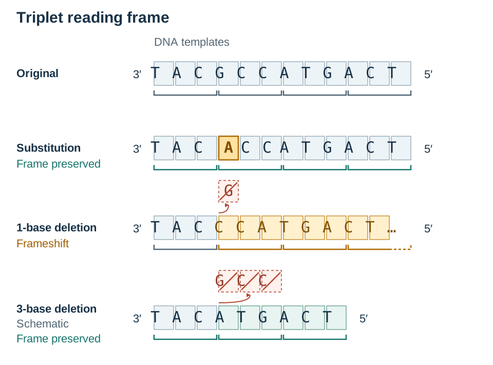
- Point mutation: A change in one or a few nucleotide bases within a single gene, typically caused by incorrect base pairing during replication.
- Substitution: One base is replaced by another. For example, in sickle cell anaemia, a single base substitution alters the codon for glutamic acid into a codon for valine. This changes one amino acid in the haemoglobin chain, causing red blood cells to deform into sickles under low oxygen. If a substitution changes a base but still codes for the same amino acid, it is a silent mutation.
- Insertion or Deletion: The addition or removal of a base changes the triplet reading frame for every codon downstream from that point (a frameshift). In cystic fibrosis, the most common mutation is a 3-base deletion in the CFTR gene, leading to the loss of a phenylalanine amino acid and producing non-functional chloride channels that cause thick mucus accumulation. Because exactly three bases are removed, the reading frame is not shifted; the protein simply lacks one amino acid (phenylalanine).
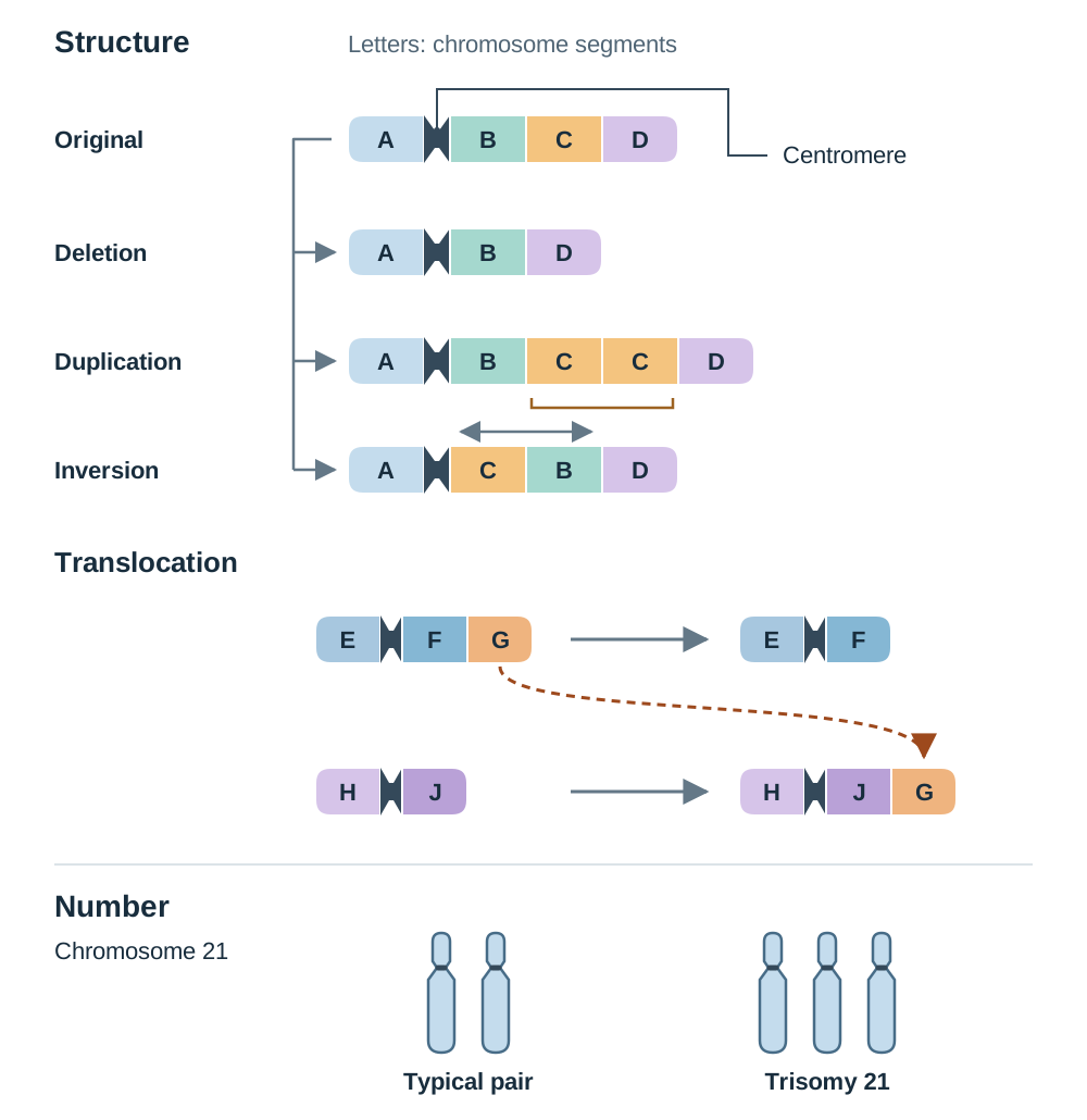
- Chromosomal mutation: A large-scale alteration in chromosome structure or chromosome number.
- Structural changes: Occur by deletion (loss of a chromosome segment, e.g. cri-du-chat syndrome on chromosome 5), duplication (a segment is repeated), inversion (a segment breaks off, flips, and rejoins), or translocation (a segment moves to a non-homologous chromosome, such as the translocation between chromosomes 9 and 22 in chronic myeloid leukaemia).
- Changes in number: Occur when chromosomes fail to separate during meiosis. Down syndrome (trisomy 21) is caused by an extra chromosome 21, resulting in 47 chromosomes instead of 46.
Cancer Biology, Factors, and Treatments
A carcinogen is any agent that causes cancer. Many carcinogens are mutagens that damage genes controlling the rate of mitosis. When these regulatory genes mutate, cells undergo abnormal regulation and divide uncontrollably, forming a tumour.
- Benign tumours: Enclosed cell masses that do not invade adjacent tissues or spread.
- Malignant tumours: Invasive cell masses that infiltrate nearby organs and can enter the bloodstream or lymphatic system to form secondary tumours elsewhere (metastasis).
Cancer Factors, Prevention, and Treatments
Cancer development is influenced by several interacting factors:
- Genetic susceptibility: Inheriting specific mutated alleles increases risk. For example, inherited mutations in BRCA1 or BRCA2 substantially raise the risk of breast and ovarian cancers, though inheriting the allele does not make cancer certain, because further mutations are still needed for cancer to develop.
- Environmental and lifestyle factors: Carcinogens such as tobacco smoke cause mutations in lung tissue. Evaluating public health statements requires looking at evidence: while smoking dramatically raises the probability of lung cancer, some non-smokers develop it and some smokers do not, reflecting individual genetic susceptibility and environmental exposure.
- Prevention: Irish public health initiatives include school-based HPV vaccination (preventing cervical and throat cancers) and national screening programmes (CervicalCheck, BreastCheck, and BowelScreen) to catch abnormal cellular changes early.
- Treatments: Surgery physically removes tumours; radiotherapy directs targeted radiation to destroy cancer cell DNA; chemotherapy uses cytotoxic drugs to kill rapidly dividing cells; and immunotherapy stimulates the body's own immune system to recognise and destroy cancer cells.
Key terms
- Gene
- A section of DNA that codes for the production of a specific protein or polypeptide.
- DNA Replication
- The semiconservative synthesis of an identical copy of a cell's DNA during interphase prior to cell division.
- Semiconservative Replication
- A mechanism of DNA replication in which each newly formed double helix consists of one intact parent strand and one newly synthesised daughter strand.
- Complementary Base Pairing
- The precise pairing of nitrogenous bases in nucleic acids via hydrogen bonds: adenine pairs with thymine (or uracil in RNA) and guanine pairs with cytosine.
- DNA Polymerase
- An enzyme that builds new DNA strands by joining complementary nucleotides together and proofreading base pairs during replication.
- RNA Polymerase
- An enzyme that binds to a promoter on DNA, unwinds the double helix, and joins complementary RNA nucleotides to synthesise an mRNA strand.
- Transcription
- The copying of a sequence of genetic information from a DNA template strand into a complementary single strand of messenger RNA (mRNA).
- Translation
- The decoding of an mRNA nucleotide sequence into a specific sequence of amino acids to form a polypeptide chain at a ribosome.
- Codon
- A sequence of three consecutive nucleotides on an mRNA molecule that codes for a specific amino acid or acts as a start or stop signal.
- Anticodon
- A triplet of nitrogenous bases on a tRNA molecule that is complementary to a specific codon on an mRNA transcript.
- Start Codon (AUG)
- The specific mRNA codon that initiates translation and specifies the amino acid methionine.
- Stop Codon
- One of three mRNA codons (UAA, UAG, UGA) that signals the termination of translation; it does not code for an amino acid.
- Peptide Bond
- The covalent chemical bond formed between adjacent amino acids in a growing polypeptide chain during translation.
- Mutation
- A change in the amount or structure of the DNA (or chromosomes) of an organism.
- Mutagen
- Any physical, chemical, or biological agent that increases the rate of mutations above the natural spontaneous level.
- Point Mutation
- A mutation that alters one or a small number of nucleotide bases within an individual gene, such as by substitution, deletion, or insertion.
- Chromosomal Mutation
- A large-scale mutation involving a change in chromosome structure (deletion, duplication, inversion, translocation) or chromosome number.
- Carcinogen
- Any agent that causes cancer, frequently by mutating genes that regulate cell division.
Check yourself
What is the sequence of the mRNA strand transcribed from the DNA template sequence AAT GCG?
UUA CGC.
Why does a single nucleotide deletion usually cause more severe damage to a protein than a single base substitution?
A deletion causes a frameshift mutation, shifting the triplet reading frame so that every codon and amino acid downstream of the mutation is altered, whereas a substitution typically affects only a single amino acid.
Why is it essential that DNA replication takes place before mitosis in the cell cycle?
Replication ensures that each chromosome is duplicated into two identical sister chromatids, so that when they separate during mitosis, both daughter cells receive a complete and identical copy of the genetic material.
Which enzyme unwinds the DNA double helix during transcription?
RNA polymerase.
Give one example of a structural chromosomal mutation and one example of a mutation in chromosome number.
Structural mutation: deletion in cri-du-chat syndrome (or translocation in chronic myeloid leukaemia). Chromosome number mutation: Down syndrome (trisomy 21).
State one limitation of using a two-dimensional 'flat ladder' diagram to model DNA.
It does not show the natural three-dimensional helical twist of the double helix (or it represents a tiny sequence of bases rather than the thousands found in a real gene).