

The breathing system enables gas exchange between the air and the bloodstream, taking in oxygen for cellular respiration and getting rid of carbon dioxide. The lungs sit inside the airtight thoracic cavity, where ventilation is driven by volume and pressure changes produced by the diaphragm and external intercostal muscles. Gaseous exchange happens across the thin, moist walls of hundreds of millions of alveoli by diffusion. Carbon dioxide level, not oxygen level, is the main factor controlling normal breathing rate. When blood carbon dioxide rises, the medulla oblongata in the brain detects the drop in pH and speeds up breathing via a negative feedback loop.
Anatomy of the Respiratory Tract
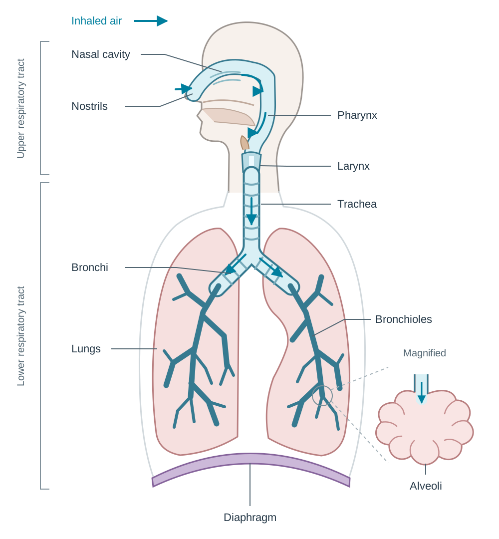
The breathing system is divided into the upper respiratory tract (nose, pharynx, and larynx, located in the head and neck) and the lower respiratory tract (trachea, bronchi, bronchioles, and lungs). The lungs sit inside the thoracic cavity (chest cavity), protected by the ribs, sternum, and vertebral column.
Air follows a precise route through the system: nostrils nasal cavity pharynx larynx trachea bronchi bronchioles alveoli.
The Upper Tract
Air enters through the nostrils into the nasal cavity, which is split down the middle by a cartilage septum. Nasal breathing conditions the incoming air in three ways:
- Filtration: Hairs in the nostrils trap large particles, while sticky mucus produced by the lining traps dust, pollen, and bacteria.
- Moistening: Water evaporates from the mucous membrane to humidify the air, preventing delicate internal exchange surfaces from drying out.
- Warming: Many capillaries just under the lining of the nose warm the air to near body temperature ().
Conditioned air then passes into the pharynx (throat), a shared muscular corridor for both air and food. At its base lies the larynx (voice box), supported by cartilage and containing two elastic vocal cords that vibrate when air passes over them to generate sound.
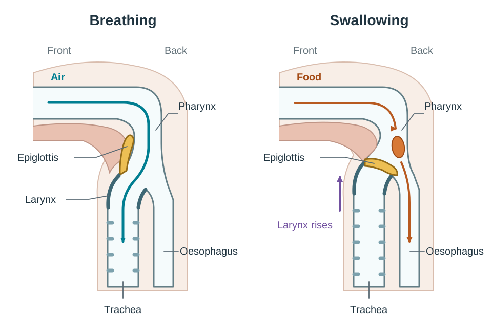
To prevent choking, a flap of flexible cartilage called the epiglottis sits above the larynx. When you swallow, the larynx moves up and the epiglottis closes over the entrance to the larynx and trachea (the glottis). This diverts swallowed food and liquid into the oesophagus behind it.
The Lower Tract
The trachea (windpipe) carries air down into the thorax. Its wall is reinforced by C-shaped rings of tough cartilage. These rings keep the trachea open during the pressure drops of inhalation. The open gap of each C-ring lies against the oesophagus, allowing swallowed food boluses to slide down without catching.
At its lower end, the trachea splits into two primary bronchi, each entering one lung. Inside the lungs, the bronchi branch repeatedly into narrower airways called bronchioles.
- Ciliated epithelium and mucus: The trachea and bronchi are lined with goblet cells and ciliated epithelial cells. Goblet cells secrete sticky mucus to trap foreign particles and pathogens. Cilia are microscopic, hair-like projections that beat in coordinated waves to sweep dirty mucus upward toward the pharynx to be swallowed and sterilised by stomach acid. Chemicals in tobacco smoke paralyse these cilia, meaning mucus collects in the airways and causes a persistent smoker's cough.
- Bronchiole smooth muscle: Unlike larger airways, the smallest bronchioles have no cartilage. Instead, their walls contain smooth muscle. In an asthma attack, these muscles contract suddenly in response to allergens, cold air, or exercise. This narrows the airways and causes breathlessness and wheezing.
Alveoli, Gaseous Exchange, and Circulation
Before looking at the lungs in detail, make sure you keep three distinct terms separate:
- Breathing (ventilation) is the physical movement of air into and out of the lungs.
- Gaseous exchange is the diffusion of oxygen from the alveoli into the blood, and of carbon dioxide from the blood into the alveoli.
- Cellular respiration is the enzymatic release of energy from glucose inside cells. It uses oxygen and produces carbon dioxide as a waste product.
Breathing and gaseous exchange supply the oxygen needed for cellular respiration and remove the carbon dioxide it generates.
Alveolar Structure and Function
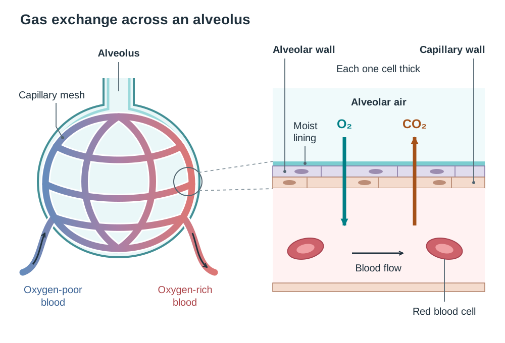
Each of the smallest bronchioles ends in a cluster of tiny air sacs called alveoli. The lungs contain about 300 to 500 million alveoli, providing a total surface area of roughly . This huge surface area allows large amounts of gas to diffuse at the same time.
Every single alveolus exhibits three crucial structural features:
- Wall one cell thick: The alveolar wall is made of a single layer of flattened squamous epithelial cells. Because the capillary wall alongside it is also just one cell thick, the total diffusion pathway is less than .
- Moist inner lining: A thin film of moisture lines each air sac. Oxygen dissolves in this moisture before diffusing across the cell membranes into the blood.
- Dense capillary mesh: A rich network of microscopic blood vessels wraps tightly around each alveolus. Continuous blood flow rapidly carries oxygen away and brings carbon dioxide in, maintaining a steep concentration gradient.
In respiratory conditions such as emphysema, long-term irritation from pollutants breaks down alveolar walls. This turns millions of tiny air sacs into fewer, larger, ragged sacs, drastically reducing surface area and leaving the person short of breath.
The Link to the Circulatory System
The breathing system works directly with the pulmonary circuit of the blood system. Deoxygenated blood leaves the right ventricle of the heart through the pulmonary artery and travels to the capillaries surrounding the alveoli. In the lungs, blood absorbs oxygen and loses carbon dioxide. The newly oxygenated blood leaves the lungs through the pulmonary vein, entering the left atrium of the heart to be pumped around the body through the systemic circuit.
Gas Transport in the Blood
- Oxygen: Most oxygen is carried inside red blood cells bound to haemoglobin as oxyhaemoglobin, with a small fraction dissolved directly in blood plasma.
- Carbon dioxide: Most carbon dioxide is converted into hydrogencarbonate ions () and carried dissolved in the blood plasma.
The Mechanics of Ventilation
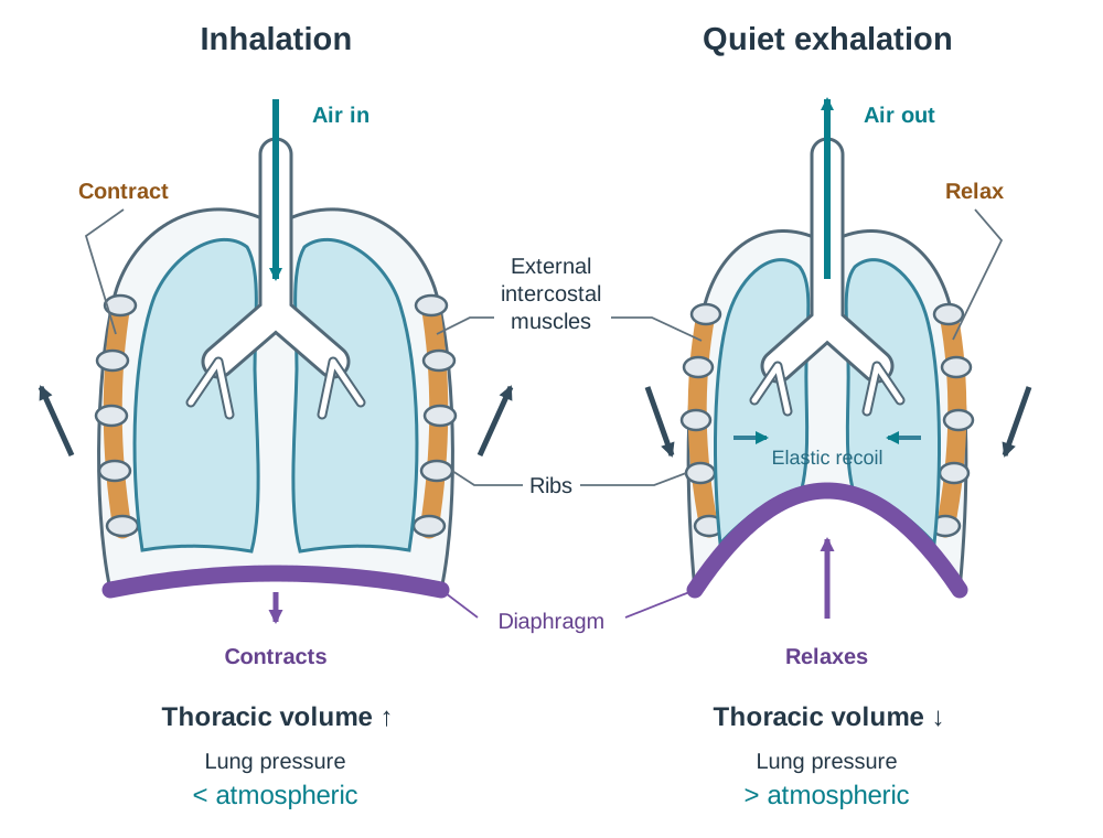
The lungs contain no muscle tissue of their own and cannot inflate or deflate themselves. Ventilation depends on altering the volume of the airtight thoracic cavity using the diaphragm and the external intercostal muscles.
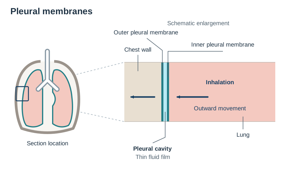
Each lung is enclosed by two pleural membranes. The outer membrane lines the inside of the chest wall, while the inner membrane covers the lung surface. Between them sits the pleural cavity, filled with a thin film of pleural fluid. This fluid acts as a lubricant to prevent friction during breathing movements. Because of surface tension, the pleural fluid also holds the lung surface tight against the chest wall. When the chest cavity expands, the lungs are pulled open with it.
| Feature | Inhalation (Active Process) | Exhalation (Normally Passive Process) |
|---|---|---|
| Nerve impulses | Impulses arrive from the medulla oblongata | Impulses from the medulla oblongata stop |
| External intercostal muscles | Contract, pulling ribs upward and outward | Relax, allowing ribs to drop downward and inward |
| Diaphragm | Contracts and flattens downward | Relaxes and curves upward into a dome shape |
| Thoracic cavity volume | Increases | Decreases |
| Pressure inside lungs | Falls below atmospheric pressure | Rises above atmospheric pressure |
| Air movement | Air is drawn into the lungs | Air is pushed out of the lungs |
Normal, quiet exhalation requires no muscular effort. The diaphragm and external intercostal muscles simply relax, and the natural elastic recoil of the stretched lung tissue pushes the air back out. During heavy exercise, however, exhalation becomes active: internal intercostals and abdominal muscles contract to pull the ribs down faster and compress the chest cavity.
Differences Between Inhaled and Exhaled Air
- Oxygen: Drops from in inhaled air to in exhaled air because oxygen diffuses into the blood.
- Carbon dioxide: Rises from in inhaled air to in exhaled air because carbon dioxide diffuses out of the blood.
- Nitrogen: Remains unchanged at because the human body does not use atmospheric nitrogen gas.
- Water vapour: Exhaled air is always saturated because moisture evaporates from the warm, wet lining of the respiratory tract.
Control of Breathing and Plant Comparison
Normal breathing is governed automatically by the respiratory centre in the medulla oblongata of the brainstem. While you can voluntarily hold your breath or breathe faster for a short time, your brain's automatic control will override your conscious effort once carbon dioxide builds up.
Negative Feedback Control of Breathing
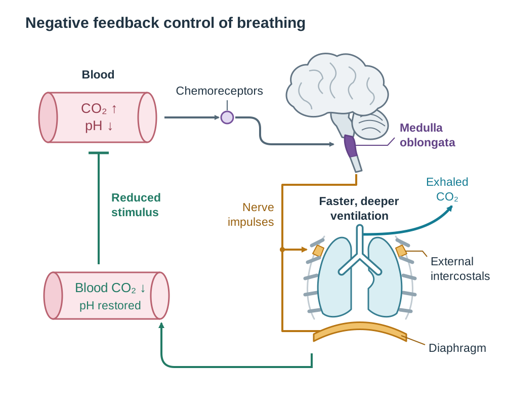
Breathing rate is regulated by homeostasis via a negative feedback mechanism that monitors blood carbon dioxide, not oxygen shortages:
- Stimulus: During exercise, active muscle cells respire faster and release more carbon dioxide into the blood.
- Chemical reaction: The carbon dioxide dissolves in the blood plasma and reacts with water to form weak carbonic acid (), which dissociates to produce hydrogen ions () and hydrogencarbonate ions ():
- Detection: The rising concentration lowers blood pH, making it slightly more acidic. Special chemoreceptors in the medulla oblongata and major arteries detect this drop in pH.
- Correction: The medulla oblongata fires more frequent nerve impulses to the diaphragm and external intercostal muscles, making breathing faster and deeper.
- Feedback: The increased ventilation expels carbon dioxide from the alveoli faster. As carbon dioxide leaves the blood, blood pH rises back to its normal level (around pH 7.4). The medulla oblongata senses this recovery and slows breathing back down to its resting rate.
Carbon Dioxide Control: Humans vs Plant Stomata
Carbon dioxide concentration is a primary controlling factor in both humans and plants, but causes contrasting physiological responses:
- In humans: High blood carbon dioxide causes faster, deeper breathing to get rid of the excess gas and prevent acidosis.
- In plants: Carbon dioxide concentration controls the opening and closing of stomata via the guard cells:
- In light: Photosynthesis uses up carbon dioxide faster than cellular respiration produces it, so internal carbon dioxide in leaf air spaces drops. Low carbon dioxide causes guard cells to absorb water, become turgid, and open the stomatal pore. This lets in more carbon dioxide for photosynthesis.
- In darkness: Photosynthesis stops, but respiration continues, so carbon dioxide accumulates inside the leaf air spaces. High internal carbon dioxide causes guard cells to lose water, become flaccid, and close the stomata, which reduces unnecessary water loss by transpiration.
Key terms
- Ventilation
- The physical movement of air into and out of the lungs (breathing).
- Gaseous exchange
- The diffusion of oxygen from the alveoli into the blood and carbon dioxide from the blood into the alveoli across a moist surface.
- Cellular respiration
- The metabolic process in which living cells break down glucose to release energy, consuming oxygen and producing carbon dioxide.
- Thoracic cavity
- The sealed, airtight chest cavity bounded by the ribs, sternum, and diaphragm that houses the lungs and heart.
- Pharynx
- The muscular throat region situated behind the mouth and nasal cavity that acts as a shared corridor for food and air.
- Epiglottis
- A flexible flap of cartilage that folds down over the glottis during swallowing to prevent food and liquid from entering the trachea.
- Larynx
- A cartilaginous structure at the top of the trachea housing the vocal cords, which vibrate to produce sound.
- Trachea
- The windpipe leading from the larynx to the bronchi, held open under negative pressure by C-shaped rings of cartilage.
- Cilia
- Microscopic, hair-like projections on respiratory epithelial cells that beat rhythmically to move mucus and trapped debris up the airways.
- Bronchioles
- Narrow, branched airways inside the lungs that lack cartilage and contain smooth muscle in their walls.
- Alveolus
- A microscopic, thin-walled, moist air sac in the lung where gaseous exchange between air and blood takes place.
- Pleural membranes
- Two serous membranes enclosing each lung and lining the thorax, separated by a fluid-filled cavity that reduces friction and couples lung expansion to chest movement.
- Diaphragm
- A dome-shaped sheet of muscle separating the thoracic and abdominal cavities that contracts and flattens during inhalation.
- External intercostal muscles
- Muscles between the ribs that contract during inhalation to pull the ribcage upward and outward.
- Pulmonary artery
- The blood vessel carrying deoxygenated blood from the right ventricle of the heart to the alveolar capillaries.
- Pulmonary vein
- The blood vessel carrying newly oxygenated blood from the alveolar capillaries to the left atrium of the heart.
- Medulla oblongata
- The part of the brainstem containing the respiratory centre, which monitors blood pH and adjusts breathing rate.
- Carbonic acid
- A weak acid (H2CO3) formed when dissolved carbon dioxide reacts with water, lowering blood pH.
- Chemoreceptors
- Sensory receptors in the medulla oblongata, aorta, and carotid arteries that detect changes in blood carbon dioxide and pH levels.
- Guard cells
- Specialised paired plant cells surrounding a stoma that change shape to open or close the pore in response to internal carbon dioxide and water status.
- Stomata
- Microscopic pores on the underside of plant leaves that permit gaseous exchange of carbon dioxide, oxygen, and water vapour.
Check yourself
What is the correct anatomical order of airways that an oxygen molecule passes through between the nostrils and an alveolus?
Nostrils → nasal cavity → pharynx → larynx → trachea → bronchi → bronchioles → alveoli.
Distinguish between breathing and cellular respiration.
Breathing is the physical movement of air into and out of the lungs (ventilation), whereas cellular respiration is the release of energy from food (glucose) inside cells.
Which blood vessel carries oxygenated blood away from the alveolar capillaries, and to which chamber of the heart does it go?
The pulmonary vein carries oxygenated blood to the left atrium of the heart.
Why are tracheal cartilage rings shaped like a 'C' instead of complete circles?
The open gap of the 'C' faces backwards against the oesophagus, allowing food boluses to slide down easily while still holding the airway open during inhalation.
How does high carbon dioxide affect human breathing compared to plant stomata?
In humans, high carbon dioxide stimulates faster, deeper breathing to remove it. In plants, high internal carbon dioxide in the leaf air spaces causes guard cells to lose water and close stomata.
What role does pleural fluid play in lung expansion during inhalation?
Surface tension in the pleural fluid holds the moist lung surface firmly against the chest wall, ensuring the lungs are pulled open whenever the thoracic cavity expands.