

The human circulatory system is a closed, double-circulation transport network driven by a muscular four-chambered heart. Blood consists of liquid plasma carrying red blood cells, white blood cells, and platelets. Together, these components deliver oxygen, nutrients, hormones, and heat to respiring cells while transporting wastes like carbon dioxide and urea to excretory organs. This guide covers blood composition and grouping, blood vessel architecture, cardiac anatomy and myogenic control, blood pressure and pulse measurement, experimental investigation of heart structure using primary and secondary data, and the integrated models linking circulation to other body systems.
Blood Composition and Functions
Blood is a liquid connective tissue made up of cellular elements suspended in a pale yellow fluid called plasma, which accounts for roughly 55% of total blood volume.
Blood Plasma
Plasma is mostly water (about 90%) containing dissolved proteins, nutrients, wastes, and mineral ions.
- Transports simple nutrients like glucose and amino acids in solution, along with urea, dissolved carbon dioxide, and hormones.
- Contains dissolved proteins, including albumin (which maintains osmotic balance), fibrinogen (essential for blood clotting), and antibodies, which are made by B lymphocytes to defend against infection.
- Absorbs heat from hard-working organs such as the liver and active muscles, circulating it to maintain an even body temperature of roughly 37 °C.
Blood Cells and Platelets
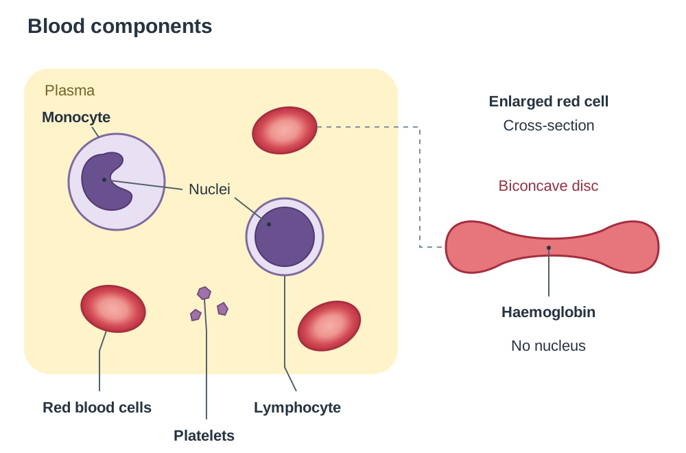
- Red Blood Cells (Erythrocytes):
- Red blood cells are biconcave discs. This shape curves inward on both surfaces, giving a large surface-area-to-volume ratio that speeds up gas exchange. Mature red cells lack a nucleus and mitochondria, leaving maximum internal space for haemoglobin and preventing the cell from using up the oxygen it carries.
- Inside the red cell, iron-rich haemoglobin binds reversibly to oxygen in the lungs to form bright red oxyhaemoglobin. When blood reaches oxygen-depleted tissues, oxyhaemoglobin breaks down and releases oxygen.
- Red blood cells are produced in red bone marrow and live for about 120 days before being broken down in the liver and spleen, where their iron is recycled.
- White Blood Cells (Leucocytes):
- White blood cells defend the body against disease. They are larger than red cells and contain a distinct nucleus.
- Monocytes are large phagocytic cells that engulf and digest pathogens and cell debris.
- Lymphocytes control specific immune responses. B lymphocytes produce antibodies, while T lymphocytes help coordinate the immune response or destroy infected cells. (Specific subtypes, such as killer cells and helper T cells, are studied in detail in the Immunity topic.)
- Platelets (Thrombocytes):
- Platelets are small, membrane-bound cell fragments that lack a nucleus.
- When a blood vessel wall is damaged, platelets stick to the exposed edges and release chemicals that convert soluble fibrinogen into insoluble strands of fibrin. This sticky mesh traps blood cells to form a clot, preventing excessive blood loss and stopping pathogens entering the body.
Blood Grouping and Rhesus Compatibility
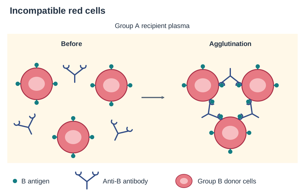
Safe blood transfusions depend on matching the surface antigen on donor red blood cells with the recipient's plasma antibody content. If incompatible blood is transfused, antibodies in the recipient's plasma bind to the foreign antigens on donor cells, causing agglutination (clumping of cells) and life-threatening complications.
The ABO System
Red cell membranes carry inherited markers called antigens, while plasma contains naturally occurring antibodies against missing antigens:
| Blood Group | Antigens on Red Cells | Antibodies in Plasma | Can Safely Receive Red Cells From | Can Safely Donate Red Cells To |
|---|---|---|---|---|
| A | A | Anti-B | Groups A, O | Groups A, AB |
| B | B | Anti-A | Groups B, O | Groups B, AB |
| AB | A and B | Neither anti-A nor anti-B | Groups A, B, AB, O | Group AB only |
| O | Neither A nor B | Both anti-A and anti-B | Group O only | Groups A, B, AB, O |
The Rhesus Factor
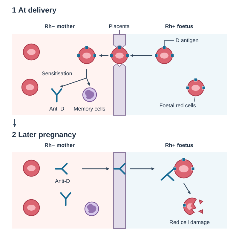
The Rhesus factor is another red blood cell antigen, known as Antigen D. A person who has this antigen is Rhesus-positive (), while someone who lacks it is Rhesus-negative ().
An individual does not naturally carry anti-D antibodies, but their immune system will produce them if exposed to blood during an incorrect transfusion or during childbirth.
When ABO and Rhesus systems are combined:
- O negative red cells have no A, B, or D antigens. Recipient antibodies have nothing to attack, making O negative the true universal donor.
- AB positive individuals have no anti-A or anti-B antibodies and already have the D antigen on their red cells, making AB positive the universal recipient.
- An patient should only receive blood to prevent sensitisation.
Haemolytic Disease of the Newborn
If an mother carries an baby, foetal red cells can leak across the placenta into maternal circulation during delivery. The mother's immune system treats Antigen D as foreign, producing anti-D antibodies and memory cells. In a later pregnancy with another baby, these maternal anti-D antibodies cross the placenta and destroy foetal red blood cells, causing severe anaemia. This is prevented by giving the mother an anti-D antibody injection within 72 hours of birth, which destroys any stray foetal red cells before her immune system becomes sensitised.
Understanding blood grouping is vital for health to ensure safe transfusions, protect pregnancies, support organ transplants, and manage blood supplies at the Irish Blood Transfusion Service.
Blood Vessels, Blood Pressure, and Pulse
Blood flows in a continuous, one-way circuit through three main types of blood vessels:
Vessel Structure and Adaptations
| Feature | Artery | Vein | Capillary |
|---|---|---|---|
| Direction of flow | Away from the heart | Towards the heart | Links arterioles to venules |
| Blood pressure | High and pulsating | Low and steady | Drops steadily along its length |
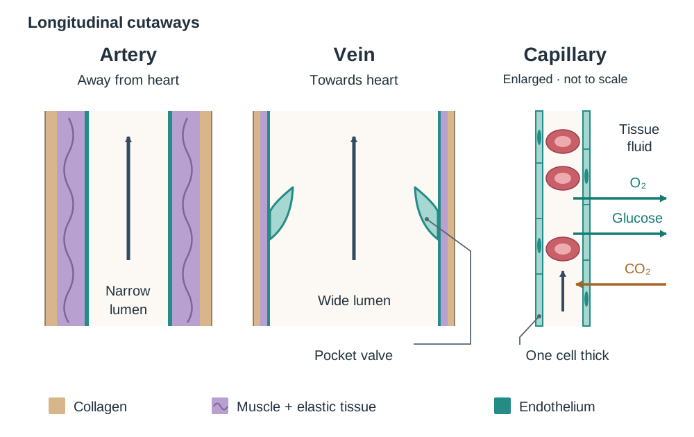
| Lumen diameter | Narrow | Wide | Microscopic (about , red cells pass single file) |
| Wall structure | Thick: tough collagen outer layer, thick muscle, and elastic fibres | Thin: little muscle and few elastic fibres | Extremely thin: single layer of endothelial cells |
| Internal valves | Absent (the semilunar valves are in the heart exits) | Present throughout limbs | Absent |
- Arteries: Pumped blood creates high pressure. Thick walls lined with collagen prevent bursting, while elastic fibres stretch during contractions and recoil between beats to keep blood moving smoothly.
- Capillaries: Their walls are just one cell thick, giving a tiny diffusion distance for rapid exchange of oxygen, glucose, and carbon dioxide between blood and tissue fluid.
- Veins: Blood pressure here is very low. Thin walls and wide lumens offer little resistance to returning blood. Veins have one-way pocket valves that close if blood starts to flow backwards. When skeletal muscles contract around veins, they compress the vessels and push blood back toward the heart.
Blood Pressure
Blood pressure is the force that blood exerts on the walls of the blood vessels. It is measured in the upper arm at the brachial artery using an inflatable cuff with a pressure gauge called a sphygmomanometer. It is recorded as two numbers in mmHg:
- Systolic pressure (the higher number): pressure in arteries when the ventricles contract.
- Diastolic pressure (the lower number): pressure in arteries when the heart relaxes.
A typical healthy resting reading for a young adult is roughly 120/80 mmHg. Persistently high blood pressure (hypertension) strains the heart muscle and damages artery linings, increasing the risk of heart attack and stroke. Contributing factors include high dietary salt, obesity, smoking, chronic stress, and lack of exercise.
Pulse Rate
Each heartbeat forces a surge of blood into the aorta, causing an elastic stretch and recoil throughout the arterial tree. This expansion is the pulse. Because each wave comes from one cardiac contraction, pulse rate equals heart rate.
You can measure pulse rate by placing two fingers over a superficial artery pressed against bone, such as the radial artery in the wrist or the carotid artery in the neck. A normal resting pulse for an adult is between 60 and 80 beats per minute (bpm). During exercise, active muscles require more oxygen and glucose for cellular respiration and produce more carbon dioxide, so heart rate rises to increase blood delivery.
Cardiac Anatomy and the Double Circulatory System
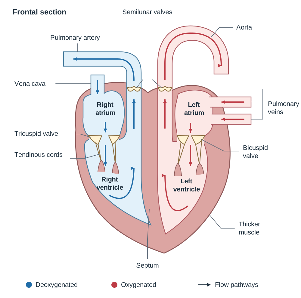
The heart is a hollow, muscular organ located in the thoracic cavity between the lungs, protected inside a fluid-filled sac called the pericardium. It consists of four distinct chambers:
Chambers and Valves
- The septum is a central muscular wall dividing the heart into right and left halves. It completely separates deoxygenated and oxygenated blood.
- Atria: The upper, thin-walled chambers. The right atrium receives deoxygenated blood from the body via the vena cava. The left atrium receives oxygenated blood from the lungs via the pulmonary veins.
- Ventricles: The lower, thick-walled chambers. The left ventricle has a muscular wall roughly three times thicker than the right ventricle. The left ventricle must pump blood under high pressure around the entire body via the aorta, whereas the right ventricle only pumps blood a short distance to the nearby lungs under low pressure.
- Atrioventricular (AV) Valves: Flaps of tissue anchored to the ventricle walls by tough tendinous cords. The tricuspid valve (three flaps) sits between the right atrium and right ventricle. The bicuspid valve (two flaps) sits between the left atrium and left ventricle. These valves close during ventricular contraction to prevent blood flowing back into the atria.
- Semilunar Valves: Half-moon-shaped pocket valves located at the exits of the ventricles, where the pulmonary artery and aorta begin. They prevent blood falling back into the ventricles when the heart relaxes.
Cardiac Muscle Tissue
The heart wall is made mainly of cardiac muscle, which shows unique structural and functional specialisations:
- It is myogenic, meaning it contracts automatically from within the tissue rather than needing nervous stimulation.
- It contracts without fatigue throughout life.
- Its cells contain abundant mitochondria to supply continuous ATP through aerobic respiration.
- Its cells are branched and interconnected, allowing electrical signals to spread across the chambers so they contract together.
Double Circulation and Portal Systems
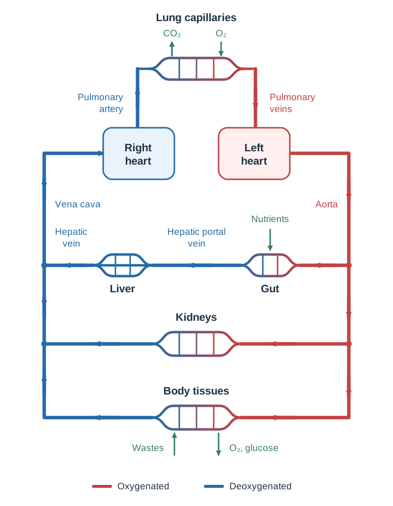
Humans have a double circulation, meaning blood passes through the heart twice during a complete journey around the body:
- Pulmonary Circuit: . This pathway carries deoxygenated blood to the lungs for gas exchange under low pressure.
- Systemic Circuit: . This pathway delivers oxygenated blood under high pressure to the rest of the body.
A portal system is a blood pathway that begins and ends in capillary beds without passing through the heart first. The hepatic portal vein carries blood loaded with absorbed nutrients from the capillary beds of the stomach and small intestine directly to the capillaries of the liver, allowing the liver to process glucose and detoxify substances before blood enters the general circulation.
Pacemaker Regulation and Cardiac Blood Supply
Although cardiac muscle generates its own rhythm, the rate and coordination of contraction are controlled by specialised pacemaker tissue.
Heartbeat Regulation
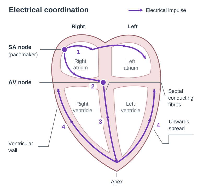
- The SA node (pacemaker), located in the upper wall of the right atrium, sends out regular electrical impulses.
- These impulses spread across both atria, causing them to contract together.
- The electrical signal reaches the AV node in the lower septum between the atria, which transmits the impulse down specialised fibres in the septum to the apex (bottom tip) of the heart.
- The impulse spreads upward through the ventricle walls, causing the ventricles to contract firmly from the bottom up, pushing blood into the arteries.
External factors adjust heart rate to match metabolic demands:
- Nervous control: When exercise increases blood carbon dioxide levels, the medulla oblongata in the brain detects this change and sends nerve impulses to speed up the SA node. When resting, different nerves signal the pacemaker to slow down.
- Hormonal control: The hormone adrenaline, released by the adrenal glands during stress, exercise, or fear, acts on the SA node to increase heart rate and pumping force.
Cardiac Blood Supply
The heart wall is too thick to absorb nutrients or oxygen directly from blood inside its chambers. Instead, the heart has its own dedicated circulation:
- Coronary arteries branch directly from the base of the aorta, spreading across the outside of the heart to deliver oxygen and glucose to cardiac muscle cells.
- Cardiac veins collect deoxygenated blood containing metabolic wastes from heart tissue and empty it into the right atrium.
If a coronary artery becomes blocked by a blood clot lodged in fatty plaque, blood flow to that section of cardiac muscle is cut off. Deprived of oxygen, that area of muscle dies, resulting in a coronary thrombosis or heart attack.
Investigating Heart Structure and Function
Dissecting a mammalian heart (such as a sheep's heart) allows students to collect primary data on cardiac anatomy and evaluate how structural features support functional roles.
Scientific Investigation Outline
- Aim / Hypothesis: To examine the internal and external structures of a mammalian heart, predicting that chamber wall thickness directly reflects the pressure required to pump blood to destination organs.
- Safety Considerations:
- Wear disposable nitrile gloves and eye protection throughout.
- Cut downward onto a dissecting board and away from fingers and body.
- Wash dissecting instruments and benches with disinfectant, and wash hands thoroughly with soap and warm water.
Primary Data Collection
- External Observations: Identify the rounded front (ventral) surface and find the diagonal groove containing coronary blood vessels. Identify the vessels at the top: the thick, elastic aorta leaving the center and the pulmonary artery in front of it. Squeeze both ventricles; the left side feels firm and muscular, while the right feels soft and compressible.
- Internal Measurements and Counts:
- Use a scalpel to make a shallow incision down the right ventricle wall. Count the flaps of the tricuspid valve (3 flaps) and observe the white tendinous cords securing them.
- Make a parallel cut down the left ventricle wall. Count the flaps of the bicuspid valve (2 flaps).
- Measure wall thickness using a millimetre ruler: in one sample heart, the left ventricle wall measured about 12 mm and the right about 4 mm.
- Cut upward into the bases of the aorta and pulmonary artery to observe the three pocket-like flaps of each semilunar valve.
Evaluating Against Secondary Data and Concluding
- Secondary Data Comparison: Published textbook measurements show that the human left ventricle wall is roughly three times thicker than the right, and mean arterial pressure in the systemic aorta (approx. 100 mmHg) is much higher than in the pulmonary artery (approx. 15 mmHg).
- Conclusion: The primary measurements showed the left wall about three times thicker; together with secondary pressure data (aorta about 100 mmHg vs pulmonary artery about 15 mmHg), this supports the prediction: the left ventricle wall is significantly thicker than the right ventricle wall. This muscular difference is an anatomical adaptation enabling the left ventricle to produce the high pressure needed to pump blood through systemic circulation, while the thinner right ventricle wall produces lower pressure suitable for delicate lung capillaries.
- Sources of Error and Limitations:
- Tissue softness makes precise ruler measurement variable.
- Hearts from a butcher are often trimmed, meaning major blood vessels might be damaged or incomplete.
- A sheep's heart differs slightly in size and vessel orientation from a human heart.
- Improvement: Take measurements at several points along the chamber wall and calculate an average, or compare results across multiple heart specimens.
Circulatory Interactions with Other Body Systems
The circulatory system functions as the body's primary transport highway, linking organ systems together to maintain cellular homeostasis:
- Digestive System: Villi in the small intestine absorb glucose, amino acids, water-soluble vitamins, and minerals directly into blood capillaries. This nutrient-rich blood travels via the hepatic portal vein to the liver for storage (such as glycogen synthesis) or processing before reaching body cells.
- Respiratory System: Pulmonary capillaries wrap closely around the alveoli in the lungs. Deoxygenated blood unloads dissolved carbon dioxide into the alveolar air and picks up oxygen, converting haemoglobin into oxyhaemoglobin for delivery to tissues.
- Urinary System: Renal arteries branch from the aorta to deliver blood containing urea, excess water, and mineral salts to nephrons in the kidneys. The kidneys filter out metabolic wastes, balance blood pH and volume, and return purified blood to the heart via the renal veins.
- Endocrine System: Endocrine glands release chemical messengers (hormones) directly into blood capillaries. Plasma carries these hormones throughout the body to target tissues.
Key terms
- Plasma
- The liquid matrix of blood, consisting of about 90% water, dissolved proteins, nutrients, wastes, and hormones.
- Haemoglobin
- An iron-containing protein in red blood cells that binds reversibly with oxygen to form oxyhaemoglobin.
- Oxyhaemoglobin
- The bright red compound formed when oxygen chemically binds to haemoglobin in areas of high oxygen concentration.
- Antigen
- A chemical marker, usually a protein or carbohydrate on a cell surface, that can trigger an immune response (antibody production) when the body recognises it as foreign.
- Antibody
- A specific protective protein produced by B lymphocytes in response to a foreign antigen.
- Agglutination
- The clumping together of red blood cells caused when antibodies bind to matching surface antigens on red cells.
- Universal Donor
- An individual of blood group O negative whose red blood cells lack A, B, and D antigens, allowing safe emergency donation to any recipient.
- Universal Recipient
- An individual of blood group AB positive who has no anti-A or anti-B antibodies and carries the D antigen, allowing them to safely receive red blood cells from any group.
- Rhesus Factor
- An inherited antigen (Antigen D) on the surface of human red blood cells used to classify blood as Rhesus-positive or Rhesus-negative.
- Blood Pressure
- The force that blood exerts on the walls of the blood vessels, recorded as systolic over diastolic pressure in mmHg.
- Pulse
- The rhythmic expansion and elastic recoil of an artery wall produced by the surge of blood pumped during each ventricular contraction.
- Double Circulation
- A circulatory system where blood passes through the heart twice during one complete circuit through separate pulmonary and systemic circuits.
- Septum
- The central muscular wall that divides the heart into right and left sides, preventing oxygenated and deoxygenated blood from mixing.
- SA Node (Pacemaker)
- A cluster of specialised cardiac muscle cells in the wall of the right atrium that generates regular electrical impulses to initiate each heartbeat.
- AV Node
- A node of specialised tissue in the lower septum that receives impulses from the atria and passes them to the ventricles.
- Coronary Arteries
- Blood vessels branching off the base of the aorta that deliver oxygenated blood and nutrients directly to the heart muscle.
- Cardiac Veins
- Vessels that collect deoxygenated blood from cardiac muscle tissue and drain it directly into the right atrium.
- Portal System
- A vascular pathway that begins and ends in capillary beds without passing directly through the heart.
- Hepatic Portal Vein
- A blood vessel that carries nutrient-rich blood from the capillary beds of the stomach and intestines directly to the capillaries of the liver.
- Semilunar Valves
- Half-moon-shaped pocket valves located at the exits of the ventricles that prevent blood flowing back into the heart when it relaxes.
Check yourself
Name two structural features of red blood cells and explain how each aids oxygen transport.
Their biconcave disc shape provides a large surface-area-to-volume ratio for rapid oxygen diffusion, and the absence of a nucleus or mitochondria leaves maximum room for haemoglobin while preventing the cell from consuming the oxygen it carries.
How does the SA node control the rhythm of a heartbeat?
The SA node acts as the heart's pacemaker by spontaneously generating regular electrical impulses that spread across the atria causing them to contract, before passing to the AV node and down the septum to trigger ventricular contraction.
What is the primary anatomical difference between the hepatic portal vein and the hepatic vein?
The hepatic portal vein carries nutrient-laden blood from the stomach and small intestine capillary beds directly to the liver capillaries, whereas the hepatic vein drains blood from the liver capillaries into the inferior vena cava.
Why is the muscular wall of the left ventricle significantly thicker than that of the right ventricle?
The left ventricle must generate high pressure to pump blood all the way around the body through the systemic circuit, whereas the right ventricle only needs to pump blood under low pressure a short distance to the lungs.
What two numbers are recorded during a blood pressure measurement, and what does each represent?
Systolic pressure is the higher number and represents the pressure in arteries when the ventricles contract; diastolic pressure is the lower number and represents the pressure in arteries when the heart relaxes.