

Every living thing is made of cells. This note covers the two main types of cells, the internal structures of plant and animal cells and what each part does, how multicellular organisms are organised, and how we use light and electron microscopes to examine them.
Levels of Organisation and Cell Theory
Cell theory forms the bedrock of biology and rests on three core principles:
- All living organisms are composed of one or more cells.
- The cell is the most basic unit of life.
- All cells arise from pre-existing cells through cell division.
In unicellular organisms such as bacteria or amoebae, a single cell carries out every metabolic, sensory, and reproductive task. Multicellular organisms, like plants and humans, rely on cellular specialisation and division of labour. Specialised cells group together to form higher levels of complexity:
Definitions and Examples of Organisational Levels
- Cell: The basic structural and functional unit of life (e.g. cardiac muscle cell).
- Tissue: A group of similar cells working together to carry out a specific function (e.g. muscle tissue, xylem).
- Organ: A distinct structure composed of several different tissues working together to carry out a particular function (e.g. the heart, a plant leaf).
- Organ System: A group of organs working together to perform a major body function (e.g. the circulatory system).
- Organism: An individual living entity capable of independent survival (e.g. a human, an oak tree).
Because different cells do different jobs, multicellular organisms can grow larger and survive with greater efficiency than single independent cells.
Prokaryotic and Eukaryotic Cells
Every cell on Earth falls into one of two structural categories based on how its genetic material is organised.
Prokaryotic Cells
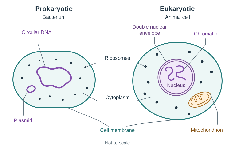
Prokaryotic cells do not have a membrane-bound nucleus or membrane-bound organelles. Their genetic material is a single, circular loop of DNA that lies free in the cytoplasm, often accompanied by tiny accessory rings of DNA called plasmids. Bacteria are the classic example. They are small, typically measuring between and across.
Eukaryotic Cells
Eukaryotic cells contain a true membrane-bound nucleus enclosing their genetic material, alongside specialized membrane-bound organelles such as mitochondria and chloroplasts. Animals, plants, fungi, and protists are all eukaryotes. Eukaryotic cells are larger, typically ranging from to across.
| Feature | Prokaryotic Cells | Eukaryotic Cells |
|---|---|---|
| Nucleus | Absent (no nuclear membrane; naked circular DNA in cytoplasm) | Present (DNA enclosed within a double nuclear membrane) |
| Membrane-bound Organelles | Absent (no mitochondria, chloroplasts, ER, or Golgi) | Present (mitochondria, chloroplasts, ER, Golgi apparatus) |
| DNA Structure | Circular loop, naked (no histones); plasmids often present | Linear chromosomes bound to histone proteins |
| Ribosomes | Present, but smaller | Present, larger |
| Cell Size | Small () | Larger () |
| Examples | Bacteria | Animal cells, plant cells, fungi, amoebae |
Using the Light Microscope and Calculating Magnification
We use microscopes to examine structures too small for the naked eye. A compound light microscope reveals microstructure (the main parts of a cell, like the nucleus, cell wall, and cytoplasm), while electron microscopes reveal ultrastructure (fine organelle details, such as mitochondrial cristae).
Parts and Functions of the Compound Light Microscope
| Part | Function |
|---|---|
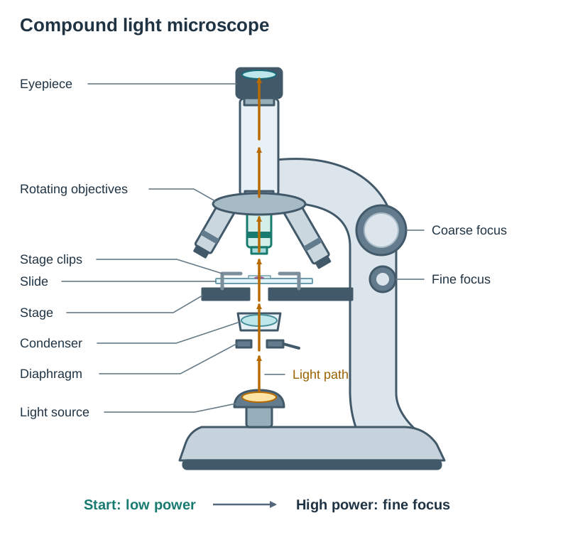
| Eyepiece lens | Magnifies the image produced by the objective lens (usually ) |
| Objective lenses | Magnify the specimen (typically low power , medium , and high power ) |
| Stage and clips | Flat platform that supports and secures the glass slide in place |
| Coarse focus knob | Moves the stage or lens rapidly over large distances to bring the specimen into rough focus |
| Fine focus knob | Moves the stage or lens very slightly to bring the image into sharp, precise focus |
| Light source / mirror | Directs light upwards through the stage aperture and specimen |
| Condenser and diaphragm | Focuses light onto the specimen and adjusts the brightness and contrast of light passing through |
Step-by-Step Method of Use
- Place the slide on the stage and secure it with the stage clips, centering the specimen over the light hole.
- Always start on the low-power objective lens. This provides the widest field of view, making the specimen easy to find.
- Looking from the side, use the coarse focus knob to bring the stage close to the objective lens. Then look through the eyepiece and slowly turn the coarse focus knob to move the stage away from the lens until the image comes into focus.
- Centre the cell you wish to view, then rotate the nosepiece to the high-power objective lens.
- Adjust only with the fine focus knob on high power so the lens does not strike and crack the glass slide.
Magnification Formula and Calculations
Total magnification on a light microscope equals the magnification of the eyepiece lens multiplied by the magnification of the objective lens:
For example, an eyepiece of combined with a high-power objective of gives a total magnification of .
When calculating the magnification of an image or drawing:
Always convert measurements to the same unit before dividing ().
Practical: Preparing and Examining Plant and Animal Cells (Outcome 1.3.3)
This practical develops primary data collection using a light microscope. Staining improves contrast across translucent cell layers, and viewing multiple fields of view makes observations repeatable and reliable.
Apparatus
Compound light microscope, clean glass slides, coverslips, mounted needle, forceps, cotton wool swabs, dropper pipettes, filter paper, iodine solution, methylene blue, and dilute disinfectant solution.
Preparing the Plant Cell Slide (Onion Epidermis)
- Use forceps to peel a thin, transparent, single-cell layer of epidermis from the inner fleshy scale of an onion.
- Spread the tissue completely flat in a small drop of water on a clean slide to stop it from wrinkling or drying out.
- Add one to two drops of iodine solution to stain the tissue.
Preparing the Animal Cell Slide (Human Cheek Epithelium)
- Gently rub the inside of your cheek with a clean cotton swab to collect epithelial cells.
- Smear the swab evenly into a drop of water or physiological saline on a clean glass slide.
- Add one to two drops of methylene blue to stain the cells.
- Immediately place the used swab into a beaker of disinfectant for safe disposal.
Mounting the Coverslip and Avoiding Air Bubbles
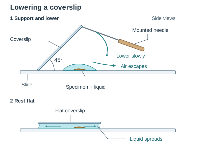
- Place one edge of the coverslip against the slide next to the liquid droplet.
- Rest the coverslip on a mounted needle and lower it slowly at a angle over the specimen.
- Lowering the coverslip at an angle pushes air out in front of the falling glass, preventing trapped air bubbles (which appear under the microscope as thick, black-bordered circles).
- Dab away excess liquid with filter paper so fluid does not get onto the microscope stage.
Why Stains Are Used
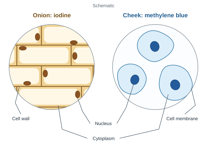
- Iodine solution stains onion cells yellow-brown. It stains the nucleus more darkly than the cytoplasm, so the nucleus, cytoplasm, and cell wall stand out clearly.
- Methylene blue stains human cheek cells blue. It binds strongly to nucleic acids, making the nucleus dark blue against a paler blue cytoplasm.
Results and Microscopic Observations
- Onion epidermal cells: Regular, elongated rectangular cells fitting together like brickwork. Visible parts include the rigid cell wall, cytoplasm, and a stained nucleus. Chloroplasts are absent because onion bulbs grow underground.
- Human cheek cells: Irregular, rounded, flexible cells seen singly or in small clumps. Visible parts include the thin cell membrane, granular cytoplasm, and a central blue nucleus. No cell wall or chloroplasts are present.
Cell Boundaries: Cell Membrane and Cell Wall
The living content of any cell is called the protoplasm, which includes both the cytoplasm and the nucleus. Two distinct boundaries can separate this living material from the outside environment.
The Cell Membrane (Plasma Membrane)
- Location: Encloses the protoplasm of all cells. In plant cells, it sits directly inside the cell wall.
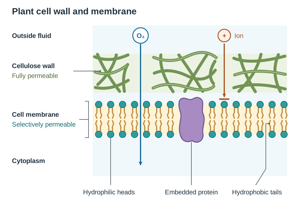
- Structure: Composed of a phospholipid bilayer with proteins embedded in it. Each phospholipid molecule consists of a polar, hydrophilic (water-attracting) phosphate head and two non-polar, hydrophobic (water-repelling) fatty acid tails. In water, they spontaneously form a bilayer: heads point outward toward the watery cytoplasm and fluid outside, while tails turn inward toward each other.
- Function: The cell membrane has selective permeability (it is semi-permeable). It allows small, uncharged molecules such as oxygen and carbon dioxide to cross freely, while controlling or blocking ions and large molecules.
- Process: Regulates osmosis, diffusion, and active transport.
The Cell Wall
- Location: Found outside the cell membrane in plants, fungi, and bacteria. Animal cells never have a cell wall.
- Structure: In plant cells, it is a tough, rigid layer made of interwoven cellulose fibres. (Fungal walls contain chitin, and bacterial walls are made of peptidoglycan, not cellulose).
- Function: The cell wall is fully permeable to water and dissolved substances. When a plant cell absorbs water by osmosis, the central vacuole swells and pushes the cytoplasm out against the wall. This outwards push is turgor pressure. The strong cellulose wall resists this pressure, keeping the cell firm (turgid) and preventing it from bursting.
- Process: Provides structural support, gives the plant cell a fixed shape, and maintains turgidity.
The Nucleus, Cytoplasm, and Energy-Transforming Organelles
The Nucleus and Nuclear Pores
- Location: Suspended in the cytoplasm of eukaryotic cells.
- Structure: Surrounded by a double nuclear membrane perforated by microscopic protein-lined nuclear pores. Inside lies the nucleolus and chromatin (DNA loosely wrapped around histone proteins). Chromatin condenses into distinct chromosomes during cell division.
- Function: The nucleus controls the cell's activities by directing protein synthesis. It holds the genetic code for almost all cellular proteins. Nuclear pores allow materials to pass between the nucleus and the cytoplasm, most notably letting messenger RNA (mRNA) exit to the ribosomes.
The Cytoplasm
- Location: The jelly-like region between the cell membrane and the nucleus.
- Structure & Function: Consists of a watery fluid (cytosol) holding the cell's organelles. It supports organelles and provides the site for vital metabolic pathways, including glycolysis (the first stage of respiration).
Mitochondria
- Location: Cytoplasm. Very active cells (such as muscle and liver cells) contain thousands of mitochondria; inactive storage cells have few.
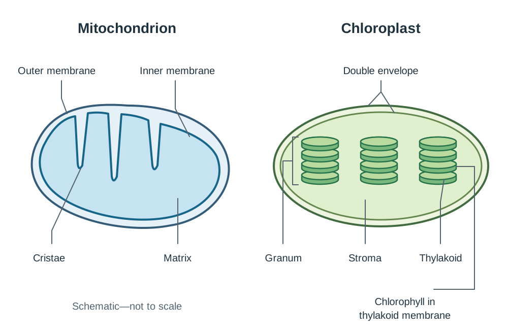
- Structure: Double-membraned organelle. The outer membrane is smooth, while the inner membrane is folded into finger-like projections called cristae. Inside lies a fluid matrix containing enzymes, ribosomes, and circular mitochondrial DNA.
- Function: Mitochondria carry out aerobic cellular respiration, releasing energy from glucose derivatives and storing it in ATP molecules. The folded cristae provide a large surface area for electron transport chains and the enzyme ATP synthase.
Chloroplasts
- Location: Found only in green plant cells exposed to sunlight (such as leaf mesophyll). They are absent in underground organs like roots.
- Structure: Double-membraned organelle containing stacks of disc-shaped thylakoids called grana, surrounded by a fluid called the stroma. Chlorophyll pigments are embedded in the thylakoid membranes.
- Function: Chloroplasts absorb light energy to drive photosynthesis, producing glucose.
Biosynthetic Organelles and Interpreting Micrographs
Ribosomes
- Structure & Location: Tiny, non-membrane-bound granules made of ribosomal RNA (rRNA) and protein. Found free in the cytoplasm or attached to the rough endoplasmic reticulum.
- Function: The site of protein synthesis. Ribosomes read mRNA transcripts and link amino acids into polypeptide chains.
Endoplasmic Reticulum (ER)
- Structure & Location: A network of folded membranes spreading through the cytoplasm from the outer nuclear envelope.
- Rough ER: Studded with ribosomes on its outer surface. It folds and transports newly made proteins.
- Smooth ER: Has no ribosomes. It synthesises lipids and phospholipids and inactivates toxins.
Golgi Apparatus
- Structure & Location: Stacks of curved, flattened membrane sacs with small round vesicles pinching off at their edges.
- Function: Receives proteins and lipids from the ER, chemically modifies them (for example, adding carbohydrate chains to make glycoproteins), and packages them into vesicles for secretion or delivery to other organelles.
Vacuole
- Structure & Location: Plant cells feature a single, permanent central vacuole bounded by a membrane and filled with cell sap (water, dissolved sugars, ions, and pigments). Animal vacuoles are small and temporary.
- Function: Pushes against the cell wall to maintain turgidity and stores water and waste products.
Identifying Organelles on Electron Micrographs
Scanning electron microscopes (SEM) provide 3D images of cell surfaces, while transmission electron microscopes (TEM) reveal thin internal cross-sections. In exam questions using secondary micrograph images, look for these distinctive visual features:
- Nucleus: Large, rounded structure enclosed by a double membrane with gaps (the nuclear pores), often with a dark nucleolus inside.
- Mitochondrion: Oval or sausage-shaped with visible internal shelves (folded cristae) crossing the matrix.
- Chloroplast: Large, disc-shaped organelle showing distinct parallel stacks of dark membranes (grana).
- Rough ER: Parallel sheets of membrane dotted with tiny, dark granules (ribosomes).
- Golgi Apparatus: Smooth, curved stacks of flattened discs surrounded by small spherical vesicles budding off the edges.
Key terms
- Prokaryotic
- Describes an organism or cell that lacks a membrane-bound nucleus and membrane-bound organelles.
- Eukaryotic
- Describes an organism or cell that possesses a membrane-bound nucleus and membrane-bound organelles.
- Tissue
- A group of similar cells working together to carry out a specific biological function.
- Organ
- A structure composed of several different tissues working together to perform a particular function.
- Organelle
- A distinct, specialised subcellular structure suspended within the cytoplasm that performs a specific metabolic function.
- Protoplasm
- All the living contents of a cell, consisting of both the cytoplasm and the nucleus.
- Cytoplasm
- The living, jelly-like material between the cell membrane and the nucleus that suspends organelles and hosts chemical reactions.
- Cell Membrane
- A selectively permeable boundary made of a phospholipid bilayer and embedded proteins that encloses the protoplasm.
- Selective Permeability
- The property of a biological membrane that allows certain substances to cross while preventing the passage of others.
- Cell Wall
- A fully permeable, rigid structural layer made of cellulose located outside the plasma membrane of plant cells.
- Turgor Pressure
- The outward pressure exerted by the fluid contents of the vacuole and cytoplasm against the cell wall in a plant cell.
- Nuclear Pores
- Small protein-lined openings in the nuclear membrane that allow the transport of substances like mRNA between the nucleus and cytoplasm.
- Mitochondrion
- A double-membrane organelle containing folded cristae that serves as the site of aerobic cellular respiration and ATP release.
- Cristae
- The internal folds of the inner mitochondrial membrane that provide a large surface area for respiratory enzymes and ATP synthase.
- Chloroplast
- A double-membrane plant organelle containing chlorophyll organised into thylakoid stacks, responsible for photosynthesis.
- Ribosome
- A minute, non-membrane-bound cellular structure composed of rRNA and protein that acts as the site of protein synthesis.
Check yourself
What biological term describes the entire living contents of a cell, comprising both the nucleus and the cytoplasm?
Protoplasm.
Why should you always begin viewing a slide under the low-power objective lens on a microscope?
It provides the widest field of view, making the specimen easy to find, and leaves plenty of room so the lens will not hit the slide.
Arrange these five terms in increasing order of complexity: organ, cell, organism, tissue, organ system.
Cell, tissue, organ, organ system, organism.
Which stain is used when preparing a temporary slide of onion epidermal cells, and what does it do?
Iodine solution; it stains the cells yellow-brown so the nucleus, cytoplasm, and cell wall can be seen clearly.
What is the function of the nuclear pores in eukaryotic cells?
They allow substances to pass between the nucleus and the cytoplasm, such as mRNA leaving the nucleus for protein synthesis.
Why does a plant cell not burst when placed into pure water?
The rigid cellulose cell wall resists the turgor pressure created as water enters the vacuole, making the cell turgid rather than bursting.
An image of a cell measures 20 mm in diameter. If its actual diameter is 40 μm, calculate the magnification.
×500 (convert 20 mm to 20,000 μm; 20,000 ÷ 40 = 500).