

In the photoelectric effect, light shining on a metal knocks electrons out of its surface. In an X-ray tube the opposite happens: fast electrons slam into a metal target and produce high-frequency radiation. X-ray production is effectively the inverse photoelectric effect, and neither process makes sense unless we treat light as discrete packets of energy called photons.
The Photoelectric Effect and Experimental Evidence
The photoelectric effect is the emission of electrons from the surface of a metal when electromagnetic radiation of a suitable frequency falls on it. We call the released electrons photoelectrons, and when they travel around an external circuit, they form a photocurrent.
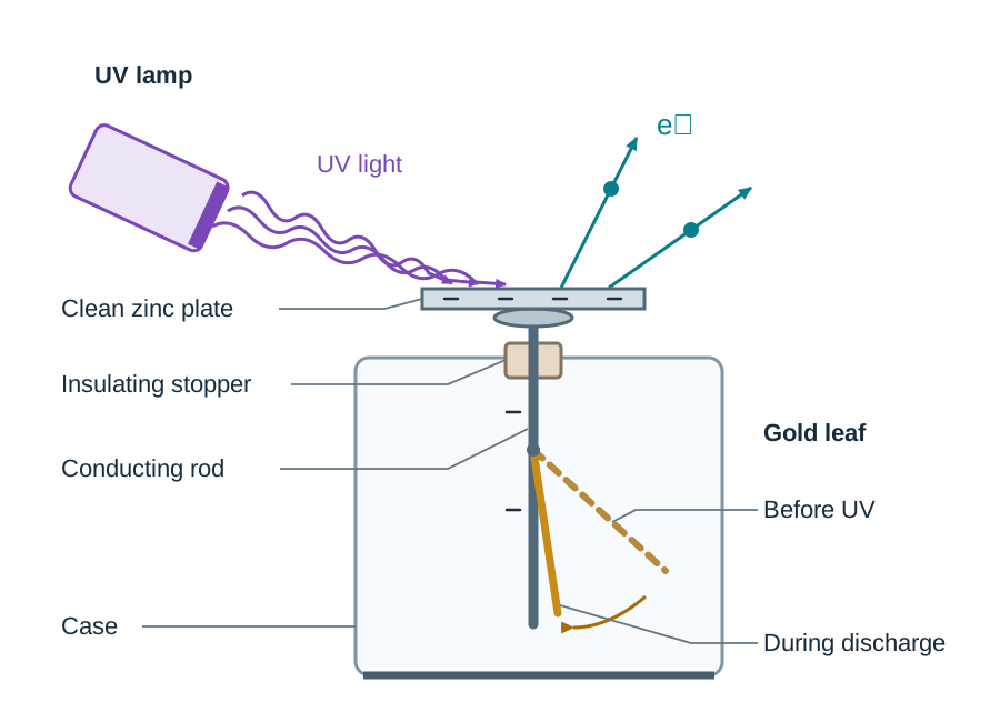
The Gold-Leaf Electroscope Demonstration
You can show this happening in the lab with a classic gold-leaf electroscope setup:
- First, rub a piece of zinc plate with emery paper or steel wool. This step is vital because it strips off the oxide layer that would otherwise insulate the metal surface.
- Place the clean zinc plate on top of the electroscope cap and give the electroscope a negative charge. The gold leaf rises because negative charges repel each other down through the rod and leaf.
- Shine ordinary visible light onto the zinc plate. Nothing happens to the leaf, even if you turn the lamp up as bright as possible.
- Now switch to an ultraviolet (UV) lamp. The leaf collapses immediately. Negative charge is leaving the plate because UV photons are knocking electrons right out of the zinc.
- Place a sheet of ordinary window glass between the UV source and the zinc plate. The leaf stops falling. Ordinary glass absorbs UV light, blocking the photons that cause emission.
- Finally, try giving the electroscope a positive charge instead. When you shine UV light on it this time, the leaf stays up. Any electron knocked loose is yanked straight back down by electrostatic attraction to the positive plate.
Frequency and Intensity: Three Standard Cases
Leaving Cert exam papers often test whether you can separate what frequency does from what intensity does:
- Case 1 (, frequency constant, intensity increasing): Increasing intensity means more photons hit the plate each second. More electrons get knocked out per second, so the photocurrent rises. The maximum kinetic energy and speed of those photoelectrons stay completely unchanged, because every photon still delivers the exact same energy pack ().
- Case 2 (, intensity constant, frequency increasing): Because intensity is constant, the same number of photons arrive each second, so the number of electrons emitted per second stays the same. But because each photon packs more energy, the maximum kinetic energy and top speed of the escaping electrons go up.
- Case 3 (, frequency constant, intensity increasing): No electrons are emitted at all. It does not matter how blinding the light is. If an individual photon does not carry enough energy () to clear the surface barrier, no electron ever escapes.
Failure of Classical Wave Theory and the Photon Model
During the nineteenth century, physicists treated light purely as a continuous wave. In that wave model, energy spread out across wavefronts and depended on wave amplitude (brightness or intensity). If that were true, classical physics made three clear predictions, and experiment proved every single one wrong:
| Wave Theory Prediction | What Actually Happens | Photon Model Explanation |
|---|---|---|
| Light of any frequency should eject electrons if the beam is made intense enough. | Below a specific threshold frequency (), no electrons ever leave the metal, no matter how bright the light. | Radiation travels in discrete packets called photons, each carrying energy . If an individual photon has , it cannot dislodge an electron. |
| Weak light should produce a measurable time lag while electrons gradually soak up wave energy. | Emission is instantaneous (taking less than a nanosecond) as soon as radiation at or above hits the surface. | An electron takes in the entire energy of one photon in a direct, one-to-one interaction; it cannot store up trickle energy from multiple low-frequency photons. |
| Higher intensity should deliver more energy to each electron, boosting electron kinetic energy. | Increasing intensity increases the number of electrons emitted per second, but leaves their maximum kinetic energy unchanged. | Intensity is simply the number of photons arriving per second. More photons free more electrons, but each photon delivers the exact same energy pack . |
In 1905, Albert Einstein solved this puzzle by proposing that light travels and interacts as localised packets of energy called photons. The energy carried by a single photon depends solely on its frequency :
where is Planck's constant () and is the speed of light in a vacuum ().
Einstein's Photoelectric Equation and Energy Conservation
Einstein looked at the collision between an incoming photon and a single electron through the lens of conservation of energy:
The work function () is the minimum energy needed to pull the most loosely bound electron free from the metal surface. At the threshold frequency (), an incoming photon provides just enough energy to free an electron with nothing left over for movement ():
Putting into Einstein's photoelectric equation gives:
Why Say 'Maximum' Kinetic Energy?
The work function applies to electrons sitting right at the metal surface. Electrons trapped deeper down collide with lattice ions on their way out, losing energy before they break free. That is why they emerge with a spread of speeds. The equation calculates the best-case escape: a surface electron that escapes clean without internal collisions, leaving with .
Graphical Analysis of versus
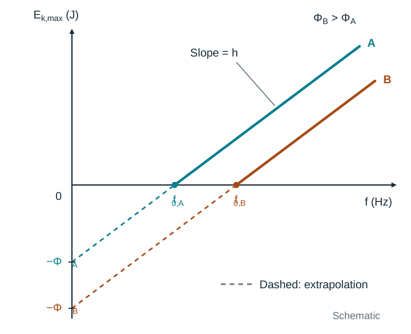
If you rearrange Einstein's equation, it matches a standard line equation:
Comparing this to , plotting maximum kinetic energy on the -axis against frequency on the -axis produces a straight line:
- Slope (): Always equals Planck's constant, .
- Horizontal intercept (-intercept): Gives the threshold frequency, , where .
- Vertical intercept (-intercept): Extrapolates back below the frequency axis to .
If you have to sketch lines for two different metals on the same set of axes, draw them with identical slopes (), but shift the line for the metal with the higher work function further to the right. Note that maximum kinetic energy is not directly proportional to frequency; it is a linear relationship with a non-zero intercept.
At atomic scale, energy is often quoted in electronvolts (eV). One electronvolt is the kinetic energy an electron gains or loses when moving across a potential difference of one volt (). Always convert electronvolts into joules before calculating speeds or working with Planck's constant.
The Photocell and Sensor Technology
A photocell is an evacuated electronic tube that uses the photoelectric effect to generate an electric current when light shines on it.
Drawing the Photocell Circuit
If an exam question asks for a diagram, draw an evacuated glass bulb containing:
- A large curved metal plate along the inside wall, labelled photocathode (low work function coating, e.g. caesium), connected to the negative terminal of a d.c. voltage supply.
- A thin central wire rod, labelled anode (collector), connected to the positive terminal.
- The outer bulb, labelled evacuated glass envelope.
- Incoming arrows directed at the curved plate, labelled incident light.
- An external circuit including a battery symbol labelled d.c. supply and a sensitive meter labelled microammeter (measures photocurrent).
Light with frequency strikes the photocathode and knocks out photoelectrons. The positive anode collects these electrons across the vacuum gap, setting up a steady photocurrent in the circuit that scales with light intensity.
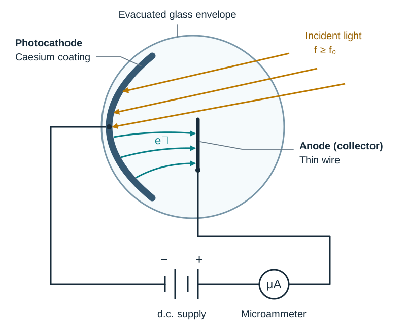
Design and Operation
- Evacuated tube: Removing air prevents escaping electrons from colliding with gas molecules, which would slow them down or scatter them off course.
- Caesium coating: Caesium has an unusually low work function, meaning visible light carries enough photon energy to trigger emission.
- Thin wire anode: Using a narrow rod ensures the collector does not cast a shadow over the photocathode.
- Emission rate and current: Because each photoelectron comes from one absorbed photon, the number of photoelectrons emitted per second matches the arrival rate of usable photons. Light intensity is the measure of incoming photons per second, so photocurrent is directly proportional to light intensity (as long as ).
From current definition and total charge , the rate of electron collection at the anode is:
Everyday Applications
- Security alarms and safety beams: A light beam shines steadily on a photocell to maintain a small current. When someone steps through the beam, the current stops, which trips an electronic relay to ring an alarm or halt industrial machinery.
- Automatic doors and street lights: People breaking light beams trigger door motors, while dusk-to-dawn sensors switch street lights on when natural daylight drops.
- Optical film soundtracks: Old cinema film prints have an optical audio track printed along the margin with alternating clear and dark bands. Light passing through the track hits a photocell, producing a fluctuating current that speakers turn back into sound.
- Smoke alarms: In optical smoke alarms, smoke particles drifting into a sensing chamber scatter light onto a photocell, generating the current pulse that sounds the buzzer.
X-Ray Production: The Hot-Cathode Tube
X-rays are high-frequency, highly penetrating electromagnetic waves discovered accidentally by Wilhelm Röntgen in 1895. In modern hospital and industrial equipment, they are produced inside an evacuated tube by firing fast electrons at a dense metal target.
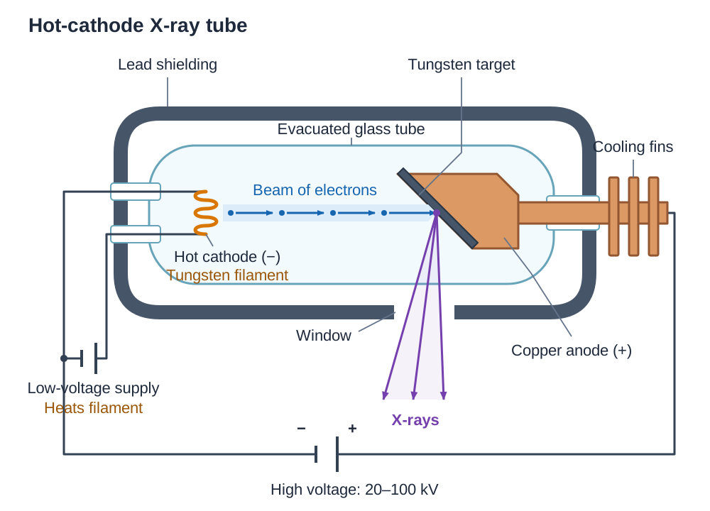
Drawing the X-Ray Tube
When sketching an X-ray tube for an exam, draw a sealed glass tube with these key parts:
- On the left, a small coiled wire labelled hot cathode (tungsten filament), wired to its own low-voltage power source labelled low-voltage supply (heats filament).
- An arrow pointing across the tube, labelled beam of electrons.
- On the right, a heavy angled metal block labelled tungsten target on copper anode, with external fins labelled cooling fins / circulating oil.
- A separate high-voltage supply between cathode and anode, labelled high voltage (20 kV – 100 kV), anode positive.
- The tube container labelled evacuated glass tube.
- Thick outer protective housing labelled lead shielding, leaving a small opening labelled window, with arrows pointing outward labelled X-rays.
How the Tube Functions
- Thermionic emission: The low-voltage circuit sends a current through the filament, heating the tungsten filament to a very high temperature (typically over ). Thermionic emission is the release of electrons from the surface of a heated metal when thermal energy allows electrons to overcome the metal's work function.
- Acceleration: An accelerating potential difference between and sets up a powerful electric field that pulls electrons away from the negative filament and drives them toward the positive anode. The vacuum allows them to cross without crashing into air molecules.
- Kinetic energy transfer: Assuming an electron begins nearly at rest, the electrical work done across the gap becomes its kinetic energy:
- Target impact: High-speed electrons slam into the tungsten target. As they decelerate violently in the strong electric fields around tungsten nuclei, they shed energy as X-ray photons.
- Heat removal: X-ray production is extremely inefficient. Less than of electron kinetic energy turns into X-rays; over turns into heat in the target. Designers use tungsten because it has a huge melting point (), and embed it in copper because copper conducts heat quickly out to cooling fins or circulating oil.
- Protective shielding: Lead absorbs stray X-rays heading in unwanted directions, leaving only a useful narrow beam that exits through the window.
Maximum Frequency and Cut-off Wavelength
The most energetic X-ray photon forms when an electron gives up all its kinetic energy in a single collision:
Tube Controls
- Accelerating voltage (): Controls the kinetic energy of the electrons, which fixes the maximum frequency () and penetrating power ('hardness') of the X-ray beam.
- Filament current (temperature): Controls the rate of thermionic emission (how many electrons leave the cathode each second), which adjusts the intensity (photon count per second) without changing individual photon energies.
Properties, Uses, and Safety
- Properties: Electromagnetic waves travelling at the speed of light in a vacuum; uncharged, so they are not deflected by electric or magnetic fields; highly penetrating; ionising; expose photographic emulsions and digital detectors.
- Uses: Medical radiography and CT scans to view broken bones or dense structures; radiotherapy to destroy cancerous tumours; security scans for luggage; non-destructive testing of metal pipe welds; X-ray crystallography to map crystal and molecular structures.
- Hazards and precautions: Because X-rays ionise atoms, they damage living cells, break chemical bonds, and trigger genetic mutations that can cause cancer. Safeguards include short exposure times, lead aprons for patients, lead-glass viewing windows for radiographers, and radiation monitoring badges.
Comparing X-Ray Production and the Photoelectric Effect
Questions asking you to compare X-ray production with the photoelectric effect are standard. To pick up full marks on a comparative question, mention both processes across similarities and differences:
| Feature | Photoelectric Effect | X-Ray Production |
|---|---|---|
| Input | Photons of electromagnetic radiation (UV or visible light). | Fast electrons accelerated through a high voltage. |
| Output | Photoelectrons released from a metal surface. | High-frequency electromagnetic radiation (X-rays) and heat. |
| Energy conversion | Photon energy turns into work function energy plus electron kinetic energy. | Electron kinetic energy turns into photon energy () and waste heat (). |
| Governing equation | ||
| Energy threshold | Minimum frequency () required to release any electrons. | Accelerating voltage () sets a maximum photon frequency () and cut-off wavelength. |
| Intensity control | Light intensity sets the number of photoelectrons emitted per second. | Filament current sets cathode temperature and the number of electrons hitting the target each second. |
| Energy control | Radiation frequency sets photoelectron kinetic energy and speed. | Accelerating voltage sets electron impact kinetic energy and X-ray penetrating power. |
| Underlying physics | One-to-one photon-electron interaction; energy is conserved. | Deceleration of electrons in nuclear fields; energy is conserved. |
How to Structure a Comparison Answer
- State the inverse relationship: In the photoelectric effect, photons strike a metal to eject electrons. In an X-ray tube, electrons strike a metal to produce photons. That is why X-ray production is called the inverse photoelectric effect.
- State a core similarity: Both processes obey the law of conservation of energy, and both prove that light exchanges energy in discrete packets (photons) rather than as continuous waves.
- State the control differences: In the photoelectric effect, light frequency sets electron kinetic energy while light intensity sets electron emission rate. In an X-ray tube, accelerating voltage sets photon energy while filament current sets X-ray beam intensity.
Key terms
- Photoelectric effect
- The emission of electrons from the surface of a metal when electromagnetic radiation of a suitable frequency falls on it.
- Photoelectron
- An electron emitted from a metal surface after absorbing energy from an incident photon.
- Photon
- A discrete packet or quantum of electromagnetic energy, given by E = hf.
- Planck's constant (h)
- The fundamental constant connecting photon energy to frequency (E = hf), equal to 6.63 × 10⁻³⁴ J s.
- Work function (Φ)
- The minimum energy needed to remove the most loosely bound electron from the surface of a metal.
- Threshold frequency (f₀)
- The minimum frequency of incident radiation below which no photoelectrons are emitted from a metal surface.
- Einstein's photoelectric equation
- The energy conservation formula for the photoelectric effect: hf = Φ + ½ m_e v_max².
- Photocurrent
- The electric current produced by the flow of photoelectrons around a circuit.
- Photocell
- An evacuated tube containing a photosensitive cathode and collector anode that converts incident light into an electric current.
- Electronvolt (eV)
- The kinetic energy gained or lost by an electron when moving through a potential difference of one volt (1 eV = 1.60 × 10⁻¹⁹ J).
- Thermionic emission
- The emission of electrons from the surface of a heated metal.
- Inverse photoelectric effect
- The conversion of electron kinetic energy into electromagnetic radiation when fast electrons decelerate at a metal target.
Check yourself
What is the threshold frequency of a metal?
The minimum frequency of incident electromagnetic radiation below which no photoelectrons are emitted from the surface of that metal.
Why does a negatively charged electroscope with a clean zinc plate discharge under ultraviolet light but not under bright red light?
Red light has a frequency below the threshold frequency of zinc, so each red photon carries less energy than the work function (hf < Φ). Ultraviolet light has a frequency above the threshold frequency, providing enough energy per photon to dislodge electrons.
Why must an X-ray tube be evacuated?
The tube is evacuated so that accelerating electrons do not collide with air molecules, which would slow and scatter them, reducing X-ray energy. The vacuum also prevents the hot filament from burning out.
What physical quantity does the slope of a graph of maximum photoelectron kinetic energy versus frequency represent?
The slope of the line equals Planck's constant, h (6.63 × 10⁻³⁴ J s).
Why is the cathode of an X-ray tube heated?
The cathode is heated by a low-voltage supply to release electrons from its surface via thermionic emission.
State two properties of X-rays that make them useful in medicine and one precaution taken because of them.
Useful properties: they are highly penetrating (passing through soft tissue but absorbed by dense bone) and they expose photographic film or digital detectors. Precaution: use lead shielding or aprons because X-rays are ionising radiation that can damage living cells and cause mutations.