

This note covers the fifteen mandatory practical investigations for Leaving Certificate Higher Level Biology. In the terminal written examination, practical questions examine core scientific principles, concise experimental steps, controls, named equipment, and precise observations at the exact level rewarded by SEC marking schemes. Every experiment is presented with exam-level methods, expected results, credited safety precautions, and past paper analysis.
Food tests for carbohydrates, protein and lipids
Aim Investigate qualitatively the presence of reducing sugars, starch, proteins, and lipids in food samples.
Apparatus and materials Glucose solution, starch suspension, albumin solution, vegetable oil, Benedict's reagent, iodine solution, Biuret reagent (sodium hydroxide and dilute copper sulfate), unglazed brown paper, deionised water, test tubes, water bath, and droppers.
Method
- Set up a negative control for each test using deionised water instead of the food sample.
- To test for reducing sugars, add Benedict's reagent to the sample and heat in a boiling water bath.
- To test for starch, add 3 drops of iodine solution to the sample.
- To test for protein, add sodium hydroxide followed by dilute copper sulfate (Biuret reagent) to the sample.
- To test for lipids, rub the sample onto unglazed brown paper and allow it to dry.
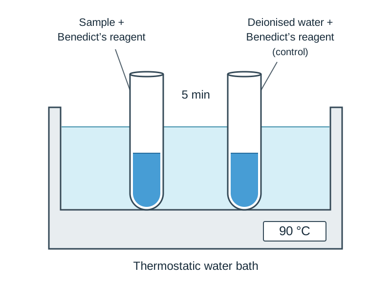
Results and calculation Benedict's reagent turns from blue to brick-red (or orange) with reducing sugars. Iodine turns from yellow-brown to blue-black with starch. Biuret reagent turns from blue to purple (or violet) with protein. Lipids form a permanent translucent spot on brown paper. All controls show no change. This investigation is qualitative so no calculation is required.
Accuracy and safety Use a water bath rather than an open flame to heat Benedict's reagent safely without splashing. Avoid skin contact with sodium hydroxide in the Biuret test because it is caustic.
How it is asked Has been asked to describe a qualitative test for a reducing sugar (add Benedict's and heat; brick-red forms). Has been asked to name the reagent and positive colour for protein (Biuret; blue to purple).
Measuring reducing sugars in food
Aim Investigate quantitatively the level of reducing sugars in food samples using a colorimeter.
Apparatus and materials Standard glucose solutions (0.1% to 1.0%), food extracts, Benedict's reagent, boiling tubes, water bath, colorimeter (620 nm), cuvettes, centrifuge, and stopclock.
Method
- Add equal volumes of Benedict's reagent to standard glucose solutions (0.1%, 0.25%, 0.5%, 1.0%) in separate boiling tubes.
- Heat all tubes simultaneously in a water bath at 95 °C for 5 minutes.
- Cool rapidly in cold water to stop the reaction, then centrifuge each tube to pellet the precipitate.
- Transfer the clear blue supernatant to a cuvette and record absorbance at 620 nm in a calibrated colorimeter.
- Repeat the procedure with the unknown food extract under identical conditions.
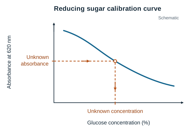
Results and calculation Plot a calibration curve with glucose concentration on the x-axis and absorbance on the y-axis. As glucose concentration increases, absorbance falls. Read the unknown sample's absorbance off the curve to find its reducing sugar concentration.
Accuracy and safety Zero the colorimeter with deionised water before readings to prevent instrumental drift. Use test-tube holders when handling hot tubes to avoid burns.
How it is asked Has been asked to distinguish between qualitative (presence or non-numerical data) and quantitative (amount or numerical data). Has not come up further in the supplied papers; a question would reasonably ask for the calibration curve or a step in the method.
Light microscope: plant cells
Aim Prepare and examine plant cells using a light microscope and relate structures to functions.
Apparatus and materials Light microscope, microscope slides, coverslips, mounted needle, forceps, onion bulb, moss leaf, iodine solution, and dropper.
Method
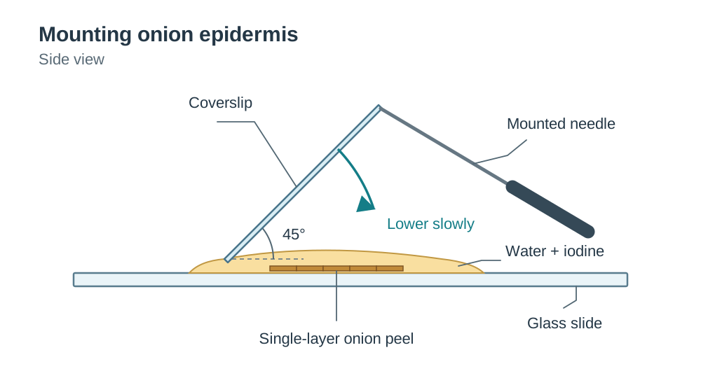
- Peel a thin, single-cell-thick layer of onion epidermis using forceps.
- Place the tissue flat on a glass slide in a drop of water.
- Add a drop of iodine stain to the tissue.
- Lower a coverslip gently at an angle using a mounted needle.
- Focus under low power using coarse focus, then switch to high power and sharpen using fine focus only.
- To see chloroplasts, mount a moss leaf in a drop of water with no stain.
Results and calculation Cell walls, cytoplasm, and stained nuclei are visible under the light microscope. Chloroplasts are visible in unstained moss leaves. Calculate actual cell size using the formula: actual size = image size ÷ magnification.
Accuracy and safety The specimen must be thin so that light can pass through it. Lower the coverslip at an angle to prevent air bubbles from being trapped.
How it is asked Has been asked how to prepare stained plant cells (cut/peel thin layer, place on slide, add iodine, add coverslip at angle). Has been asked why a thin layer is needed, and to calculate actual cell size.
Light microscope: animal cells
Aim Prepare and examine animal cells using a light microscope and relate structures to functions.
Apparatus and materials Light microscope, microscope slides, coverslips, sterile cotton swab, mounted needle, methylene blue stain, dropper, and disinfectant.
Method
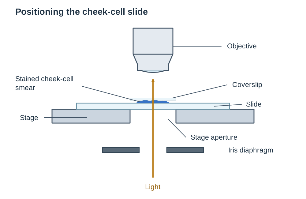
- Scrape the inside of the cheek gently with a sterile cotton swab and smear onto a slide.
- Dispose of the swab immediately into disinfectant.
- Add a drop of methylene blue stain to the cells.
- Lower a coverslip slowly at an angle using a mounted needle.
- Focus under low power using coarse focus.
- Switch to high power (x40 objective) and focus using the fine focus knob only.
Results and calculation Irregularly shaped cells with a cell membrane, cytoplasm, and darkly stained nucleus are visible. Calculate total magnification using: total magnification = eyepiece magnification × objective magnification.
Accuracy and safety Dispose of swabs in disinfectant immediately to prevent biohazard contamination. Lower the coverslip slowly at an angle to prevent trapping air bubbles and protect the lens.
How it is asked Has been asked how to prepare an animal cell slide (add stain/water to sample, lower coverslip slowly/at an angle). Has been asked why methylene blue is used (makes cells more visible) and how to view at x400 from x100 (select high-power lens, use fine focus).
Factors affecting enzyme activity
Aim Investigate factors affecting the rate of enzyme activity using catalase.
Apparatus and materials Celery extract (catalase), hydrogen peroxide, pH buffer solutions, washing-up liquid, graduated cylinders, water baths, thermometer, and stopclock.
Method
- Add pH buffer 7, a drop of washing-up liquid, and celery extract to a graduated cylinder.
- Place the cylinder and hydrogen peroxide in a water bath to reach the test temperature.
- Add hydrogen peroxide to the cylinder and start the stopclock immediately.
- Record the volume of foam produced after 2 minutes.
- Repeat at different temperatures (0–60 °C) keeping pH constant, or repeat across pH buffers (pH 4, 7, 9) keeping temperature constant in a water bath.
- Repeat using celery extract that has been boiled and cooled as a control, with all other volumes and conditions the same.
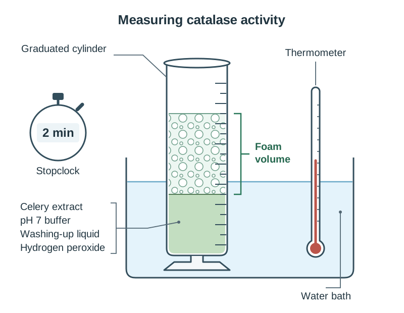
Results and calculation Rate of enzyme activity = volume of foam ÷ time. Rate increases with temperature up to an optimum (25–30 °C), then drops to zero as the enzyme denatures. Rate against pH gives a bell-shaped curve peaking at optimum pH 7. The boiled extract produces no foam, while the fresh extract produces foam, showing the enzyme was denatured.
Accuracy and safety Equilibrate enzyme and substrate in the water bath before mixing to ensure the reaction occurs at the target temperature. Dilute hydrogen peroxide is an irritant; avoid skin contact.
How it is asked Has been asked to name the enzyme (catalase), substrate (hydrogen peroxide), and products (water and oxygen). Has been asked how pH was varied (different buffers), how temperature was kept constant (water bath), how rate was measured (volume of foam per unit time), and to draw the rate curve. Has been asked how the enzyme was treated in the test (boiled) and in the control (not boiled) and to compare the results (no activity versus activity).
Factors affecting the rate of photosynthesis
Aim Investigate the effect of light intensity or carbon dioxide concentration on the rate of photosynthesis.
Apparatus and materials Elodea (pondweed), sodium hydrogencarbonate solutions, boiling tube, large beaker (water bath), lamp, metre stick, thermometer, and stopclock.
Method
- Place a cut sprig of Elodea into a boiling tube containing sodium hydrogencarbonate solution, cut stem pointing up.
- Place the tube into a large water bath at 20 °C to maintain constant temperature.
- Position a lamp at a set distance measured with a metre stick and allow 5 minutes to settle.
- Count the number of bubbles released per minute.
- Repeat at different lamp distances keeping carbon dioxide constant, or vary sodium hydrogencarbonate concentration at a fixed lamp distance.
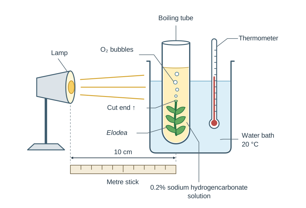
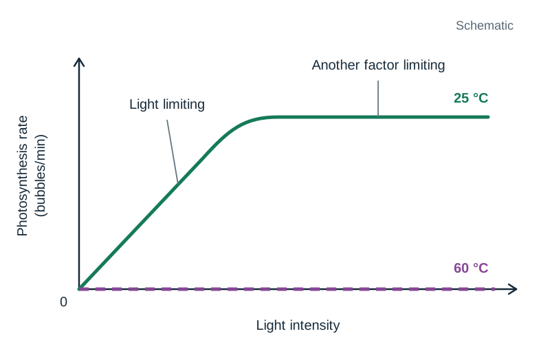
Results and calculation Rate of photosynthesis is measured as bubbles per minute. As light intensity or carbon dioxide concentration increases, the rate increases and then levels off. At 60 °C, the rate remains at zero because enzymes are denatured.
Accuracy and safety A large water bath acts as a heat shield, keeping temperature constant as lamp distance changes. Keep electrical lamp plugs dry and away from water.
How it is asked Has been asked why Elodea is suitable (aquatic plant, bubbles can be seen/counted). Has been asked how temperature was kept constant (water bath), how rate was measured (bubbles per minute), and to describe the graph (increases, then levels off).
Conditions needed for fermentation
Aim Investigate conditions necessary for fermentation and test for alcohol production.
Apparatus and materials Yeast, glucose solution, boiled deionised water, liquid paraffin, conical flasks, fermentation locks with limewater, potassium iodide, sodium hypochlorite, and water baths.
Method
- Boil glucose solution to remove dissolved oxygen, then cool.
- Add yeast to the glucose solution in a conical flask.
- Layer liquid paraffin over the mixture (or fit a fermentation lock) to maintain anaerobic conditions.
- Place in a water bath at 30 °C until bubbling stops.
- Set up controls: one flask without glucose, and one using boiled (dead) yeast.
- Filter the solution and test for alcohol by adding potassium iodide and sodium hypochlorite, then heating gently.
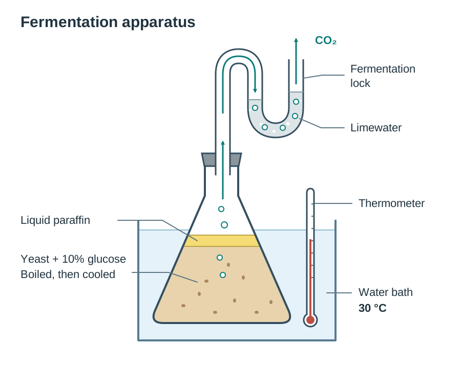
Results and calculation Carbon dioxide bubbles turn limewater milky. Fermentation stops when bubbles cease. Alcohol presence is confirmed by the iodoform test forming yellow crystals. Boiled yeast and flasks without glucose show no fermentation.
Accuracy and safety Cool the boiled glucose before adding yeast so enzymes are not denatured. Liquid paraffin excludes oxygen to maintain anaerobic conditions.
How it is asked Has been asked how anaerobic conditions were maintained (oil on top or fermentation lock). Has been asked why fermentation stops (glucose used up or alcohol toxicity), how to know it has stopped (bubbles stop), and the test for alcohol (iodoform; yellow).
Insect-pollinated and wind-pollinated flowers
Aim Investigate and compare the structural adaptations of insect-pollinated and wind-pollinated flowers.
Apparatus and materials Insect-pollinated flower (e.g. buttercup), wind-pollinated flower (e.g. grass), hand lens, dissecting microscope, scalpel, forceps, white tile, slides, and coverslips.
Method
- Place an insect-pollinated flower on a white tile and observe petals, nectaries, stamens, and carpels with a hand lens.
- Note whether petals are large and brightly coloured, and if anthers and sticky stigmas are enclosed within the flower.
- Place a wind-pollinated flower on the tile and examine under a dissecting microscope.
- Note small, dull green bracts, lack of scent, long dangling filaments with exposed anthers, and large, feathery stigmas.
- Mount pollen grains from both flowers on glass slides and examine under low power.
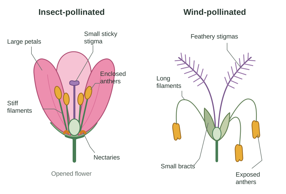
Results and calculation Insect-pollinated flowers have large coloured petals, scent, nectaries, internal stamens, small sticky stigmas, and spiky pollen. Wind-pollinated flowers have small green bracts, no scent or nectar, feathery hanging stigmas, dangling anthers, and light, smooth pollen.
Accuracy and safety Dissect fresh, mature flowers so anthers and stigmas are fully developed. Cut away from the body on a firm tile when using a scalpel.
How it is asked Has not come up in the supplied papers; a question would reasonably ask for a labelled diagram, a structural comparison table, or an adaptation related to wind or insect pollination.
Factors affecting the rate of osmosis
Aim Investigate factors affecting the rate of osmosis using a semi-permeable membrane.
Apparatus and materials Visking tubing, sucrose solutions (10%, 20%, 30%), deionised water, beakers, electronic balance, thread, scissors, and paper towels.
Method
- Soak lengths of Visking tubing in water and tie a secure knot at one end of each.
- Fill one tube with 10% sucrose and others with 20% and 30% sucrose, tying the tops securely.
- Set up a control tube filled with deionised water.
- Rinse and thoroughly blot the outside of each tube dry, then record initial masses on a balance.
- Place each tube into a beaker of deionised water for 60 minutes.
- Remove each tube, blot completely dry, and record final masses.
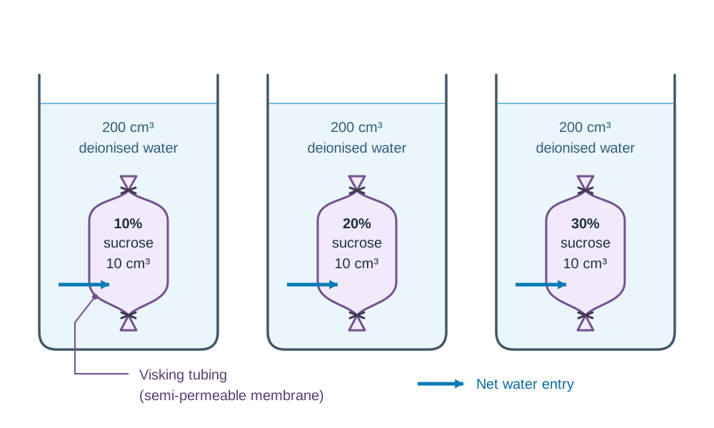
Results and calculation Percentage change in mass = [(final mass − initial mass) ÷ initial mass] × 100. Tubing containing sucrose gains mass and becomes turgid as water enters by osmosis from high to low water concentration. Steeper sucrose gradients cause faster mass gain. The water control shows no mass change.
Accuracy and safety Blot the outside of the tubing thoroughly before each weighing so adhering water does not cause errors. Tie knots tightly to prevent leakage.
How it is asked Has been asked why Visking tubing is used (acts as a selectively permeable membrane). Has been asked how osmosis was demonstrated (mass increased / tubing got fuller), and to explain mass changes of cells in sugar solutions.
Structures of the heart
Aim Dissect, display, and identify the internal and external structures of a mammalian heart.
Apparatus and materials Fresh sheep's heart, dissecting board, scalpel, scissors, seeker, and disposable gloves.
Method
- Identify the front (ventral) surface by locating the diagonal coronary artery groove.
- Squeeze both sides to identify the left side, which is firmer and thicker than the right.
- Make a shallow cut with a scalpel down the right ventricle and into the right atrium to expose the tricuspid valve.
- Make a cut down the left ventricle and into the left atrium to expose the bicuspid valve.
- Cut open the base of the aorta and pulmonary artery to expose the semilunar valves.
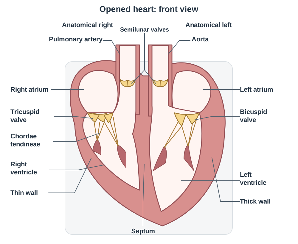
Results and calculation The left ventricular muscular wall is much thicker than the right ventricular wall. The tricuspid valve lies between the right atrium and right ventricle; the bicuspid valve lies between the left atrium and left ventricle. Semilunar valves are located at the base of the aorta and pulmonary artery.
Accuracy and safety Always cut downwards onto the dissecting board away from fingers using a scalpel. Wear gloves and wash hands with disinfectant after handling raw animal tissue.
How it is asked Has been asked how to distinguish front from back (coronary artery on front), left from right (left side thicker/firmer), instrument used to cut (scalpel), exact location of tricuspid and semilunar valves, and to compare left and right ventricular wall thickness.
Factors affecting the rate of transpiration
Aim Investigate factors affecting the rate of transpiration using a potometer.
Apparatus and materials Leafy shoot, potometer, capillary tube with scale, water reservoir with tap, beaker, petroleum jelly, fan, lamp, and stopclock.
Method
- Cut a leafy shoot under water at a slant and fit it into the potometer stopper under water.
- Seal all joints and the stopper with petroleum jelly to make the apparatus airtight.
- Clamp the potometer upright and introduce an air bubble into the capillary tube.
- Measure the distance moved by the air bubble along the scale in 10 minutes.
- Reset the bubble using the reservoir tap.
- Vary one factor (e.g. use a fan for air movement, or alter lamp distance for light), keeping all other factors constant.
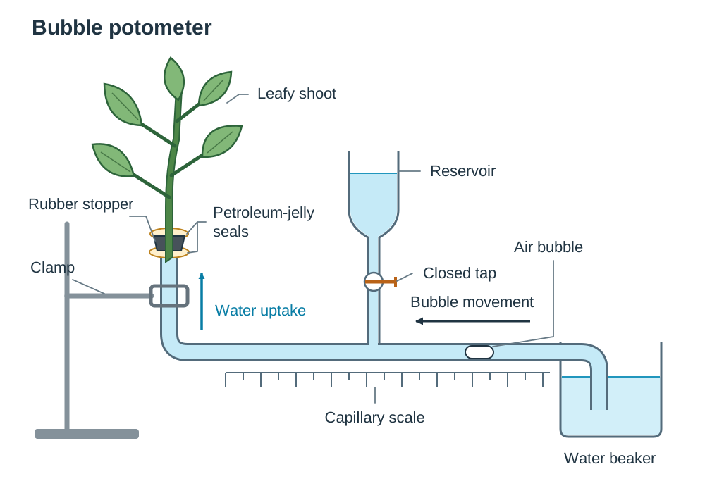
Results and calculation Transpiration rate = distance bubble moved (mm) ÷ time (min). Increased wind speed, higher light intensity, or increased temperature increases the rate of transpiration.
Accuracy and safety Cut the shoot under water to prevent air bubbles entering and blocking xylem vessels. Smear petroleum jelly on joints to ensure the system is completely airtight.
How it is asked Has not come up in the supplied papers; a question would reasonably ask why the shoot is cut under water, how airtight seals are maintained, how the bubble is reset, or the effect of a named factor on the rate.
Fieldwork: surveying a habitat
Aim Model a habitat by conducting qualitative and quantitative surveys of flora and fauna.
Apparatus and materials Frame quadrat, gridded quadrat, measuring tapes, random number table, pooter, pitfall traps, sweep net, beating tray, and identification keys.
Method
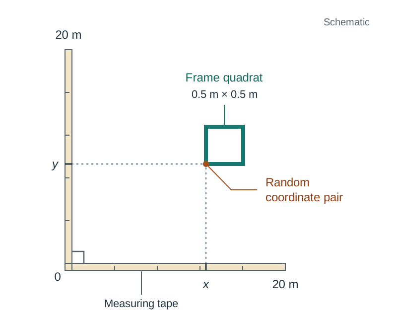
- Lay two measuring tapes at right angles to form coordinate axes across the habitat.
- Obtain pairs of random coordinates from a random number table to place quadrats without bias.
- In each frame quadrat, identify plant species using a key and record presence to calculate percentage frequency.
- Use a gridded quadrat to determine percentage cover by counting squares more than half covered by a named plant.
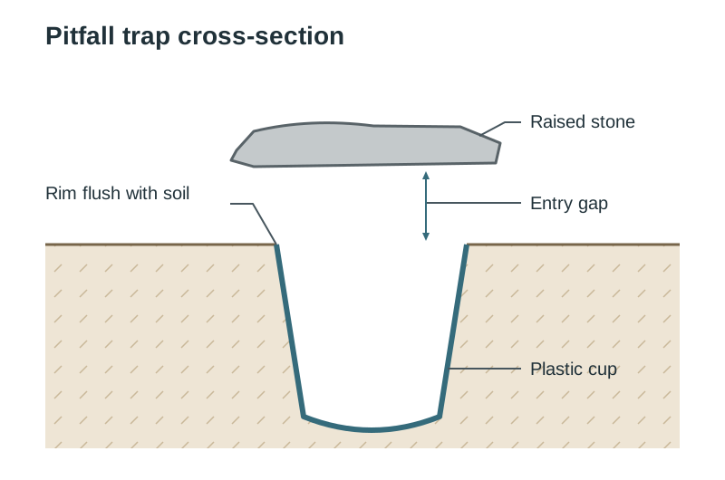
- Collect ground animals using pitfall traps (cups sunk flush into the ground left for 24 hours).
- Collect animals from vegetation using sweep nets, beating trays, and pooters.
Results and calculation Percentage frequency = (quadrats containing species ÷ total quadrats) × 100. Percentage cover is the average number of grid squares occupied. Animals collected are identified, counted, and released.
Accuracy and safety Use random coordinates rather than throwing the quadrat to prevent investigator bias. Wash hands thoroughly after field sampling to avoid contamination with soil pathogens.
How it is asked Has been asked how to obtain percentage frequency or percentage cover using a quadrat (random coordinates, count/estimate, repeat, calculate). Has been asked to name and describe animal collection apparatus (pooter: suck through tube; pitfall trap: cup flush in ground).
Fieldwork: abiotic factors and the distribution of a species
Aim Investigate quantitatively the effect of abiotic factors on the distribution and abundance of a species.
Apparatus and materials Line transect tape, frame quadrat, soil thermometer, digital light meter, soil pH meter, and clinometer.
Method
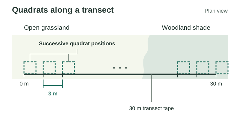
- Lay a line transect tape across an environmental gradient (e.g. from open grassland into woodland).
- Place a quadrat at regular intervals along the transect tape.
- In each quadrat, record the percentage cover or abundance of a named plant species.
- Measure light intensity at ground level inside each quadrat using a light meter.
- Insert a soil thermometer 5 cm into the ground to measure soil temperature.
- Measure soil pH using a pH meter, and slope using a clinometer.
Results and calculation Record abiotic measurements and plant abundance at each transect station. Plot transect distance on the x-axis, plant abundance on one y-axis, and the abiotic factor on the other y-axis to show correlation between factor and distribution.
Accuracy and safety Take all light readings within a short time window so changing sunlight does not alter values. Wear sturdy footwear to avoid slips and falls on uneven ground.
How it is asked Has been asked to name two abiotic factors and state how each was measured (e.g. temperature using a thermometer; light intensity using a light meter). Has been asked how quantitative plant studies were conducted along transects.
Fieldwork: feeding and symbiotic relationships
Aim Investigate and explain feeding and symbiotic relationships between organisms in an ecosystem.
Apparatus and materials Identification keys, notebook, and hand lens.
Method
- Survey the habitat to identify producers, herbivores, carnivores, and decomposers using keys.
- Observe feeding interactions directly or inspect plants for evidence of herbivory.
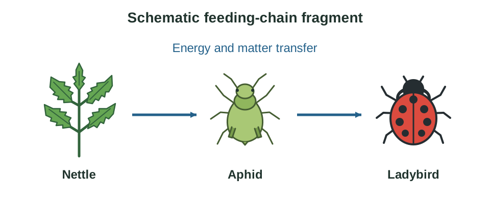
- Construct a grazing food chain of at least four named organisms starting with a producer.
- Combine multiple food chains to construct a food web for the ecosystem.
- Identify mutualism by examining clover root nodules for nitrogen-fixing bacteria (Rhizobium).
- Identify parasitism by locating aphids on plant stems or bracket fungi on trees.
Results and calculation Construct a food chain and web; arrows point from the organism eaten to the organism eating it, showing the pathway of energy transfer. Symbiosis is a close relationship between different species: mutualism benefits both, whereas parasitism benefits the parasite and harms the host.
Accuracy and safety Confirm feeding links across repeated observations rather than assuming relationships from single sightings. Cover skin cuts and wash hands after handling vegetation or soil.
How it is asked Has not come up in the supplied practical papers; a question would reasonably ask to construct a named food chain or food web, to explain the direction of energy arrows, or to define mutualism and parasitism with examples.
Factors affecting the growth of microorganisms
Aim Investigate factors affecting the growth of microorganisms using nutrient agar plates.
Apparatus and materials Nutrient agar plates, bacterial culture (Bacillus subtilis), sterile spreader, sterile paper discs, antiseptics or antibiotics, sterile forceps, incubator (25 °C), Bunsen burner, and autoclave.
Method
- Wipe the bench with disinfectant and light a Bunsen burner to maintain an aseptic work area.
- Transfer bacterial culture onto nutrient agar and spread evenly using a sterile spreader.
- Dip sterile paper discs into different antiseptics or antibiotic solutions.
- Place a disc soaked in sterile water on the plate as a negative control.
- Using flamed, cooled forceps, place each disc onto the agar surface.
- Invert the plate, secure with two small strips of tape, and incubate at 25 °C for 48 hours.
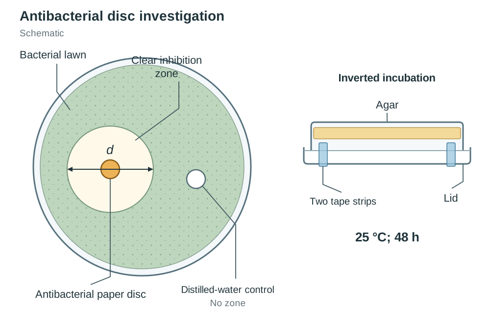
Results and calculation Bacteria grow to form a cloudy lawn. Effective antibacterial chemicals produce a clear zone of inhibition around the disc where no bacteria grow. Zone diameter is measured using a ruler. The water control disc shows no clear zone.
Accuracy and safety Do not seal the Petri dish completely, as anaerobic conditions can encourage dangerous pathogenic bacteria. Incubate at 25 °C, never 37 °C, to avoid growing human pathogens. Autoclave plates before disposal.
How it is asked Has been asked to explain sterility (free from all microorganisms) and asepsis (free from pathogens), describe two aseptic techniques (wipe bench with disinfectant, flame equipment), state the control, and give disposal method (autoclave / soak in disinfectant).
Key terms
- Hypothesis
- A testable statement or proposed explanation for an observation.
- Control
- A standard setup used as a comparison to the experiment to show the effect of the variable.
- Replicate
- A repeat of an experiment to increase the reliability or validity of data.
- Qualitative
- An observation or test indicating the presence or absence of a substance, yielding non-numerical data.
- Quantitative
- A measurement or count indicating the amount of a substance or number of organisms, yielding numerical data.
- Denaturation
- The irreversible loss of an enzyme's three-dimensional shape and function, destroying its active site.
- Osmosis
- The movement of water from a region of higher water concentration to a region of lower water concentration across a selectively permeable membrane.
- Turgor
- The pressure of the cell contents against the cell wall in a plant cell.
- Plasmolysis
- The shrinking of plant cell cytoplasm and cell membrane away from the cell wall due to water loss by osmosis.
- Transpiration
- The loss of water vapour from the leaves and aerial parts of a plant.
- Ecosystem
- A community of organisms interacting with each other and their environment.
- Abiotic factor
- A non-living physical or chemical factor that influences an ecosystem.
- Biotic factor
- A living factor that influences organisms in an ecosystem.
- Sterility
- The condition of being completely free from all living microorganisms.
- Asepsis
- The condition of being free from pathogenic or disease-causing microorganisms.
- Symbiosis
- A close relationship between two organisms of different species where at least one benefits.
Check yourself
What reagent is used to test for protein and what is the positive colour change?
Biuret reagent (or sodium hydroxide and copper sulfate); it turns from blue to purple (or violet).
Why must plant or animal tissue on a microscope slide be very thin?
To allow light to pass through the specimen so cells can be seen clearly.
Why is a water bath used when investigating the effect of pH on enzyme activity?
To keep temperature constant so there is only one variable.
What observable feature identifies the front (ventral) surface of a mammalian heart?
The coronary artery runs diagonally across the front surface.
How do you establish anaerobic conditions when preparing alcohol using yeast?
Layer liquid paraffin (or oil) over the mixture or use a fermentation lock.
What is the difference between asepsis and sterility?
Asepsis means free from pathogens; sterility means free from all microorganisms.
How is a gridded quadrat used to measure the percentage cover of a plant species?
Count the number of squares where the named plant is present or covers more than half the square, repeat, and calculate the average.