
Cells are the basic building blocks of all living things, studied in detail using a light microscope. In the Junior Cycle exam, you need to identify cell parts, describe their functions, compare plant and animal cells, and explain slide preparation practicals.
The Light Microscope and Magnification
A microscope is used because cells are far too small to be seen with the naked eye; it magnifies them so the parts can be seen.
- Eyepiece lens: The lens you look through at the top, usually magnifying 10×.
- Objective lenses: Magnify the specimen by different amounts, typically 4×, 10×, and 40×.
- Stage: The flat platform that holds the glass slide firmly in place.
- Focus knobs: The coarse focus brings the image into rough focus, while the fine focus sharpens the image.
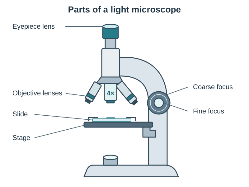
To view a slide safely, start with the lowest-power objective lens (4×) in place. Look from the side while raising the stage close to the lens. Then look through the eyepiece and turn the coarse focus knob to lower the stage slowly, increasing the gap between slide and lens, until the cells come into focus. Use the fine focus to sharpen the image.
Always multiply the lens powers together. For example, a 10× eyepiece and a 40× objective give a total magnification of 400× ().
Animal Cell Structure and Function
Animal cells contain specialised internal structures called organelles. Each organelle carries out a specific job to keep the cell alive.
- Cell membrane: A thin outer layer that controls what enters and leaves the cell. It is selectively permeable, meaning it lets some substances through but not others.
- Cytoplasm: A watery jelly where most cellular chemical reactions take place.
- Nucleus: The control centre of the cell that contains DNA and directs cell activities.
- Mitochondria: Tiny structures where cellular respiration occurs to release energy from glucose.
Under a school microscope you can see the cell membrane, cytoplasm, and stained nucleus; mitochondria are too small to see clearly.
Plant Cell Structure and Comparisons
Plant cells have the same basic parts as animal cells, along with three extra structures (remember the letters VCW):
- Vacuole: A large fluid-filled space containing cell sap that stores nutrients and keeps the plant cell firm.
- Chloroplast: A green structure containing chlorophyll that absorbs light energy to make food during photosynthesis.
- Cell wall: A tough outer layer made of cellulose that supports and strengthens the plant cell.
| Structure | Animal Cell | Plant Cell |
|---|---|---|
| Cell wall | Absent | Present |
| Chloroplasts | Absent | Present (in green tissue) |
| Large vacuole | Absent | Present |
| Cell membrane | Present | Present (inside cell wall) |
| Cytoplasm | Present | Present |
| Nucleus | Present | Present |
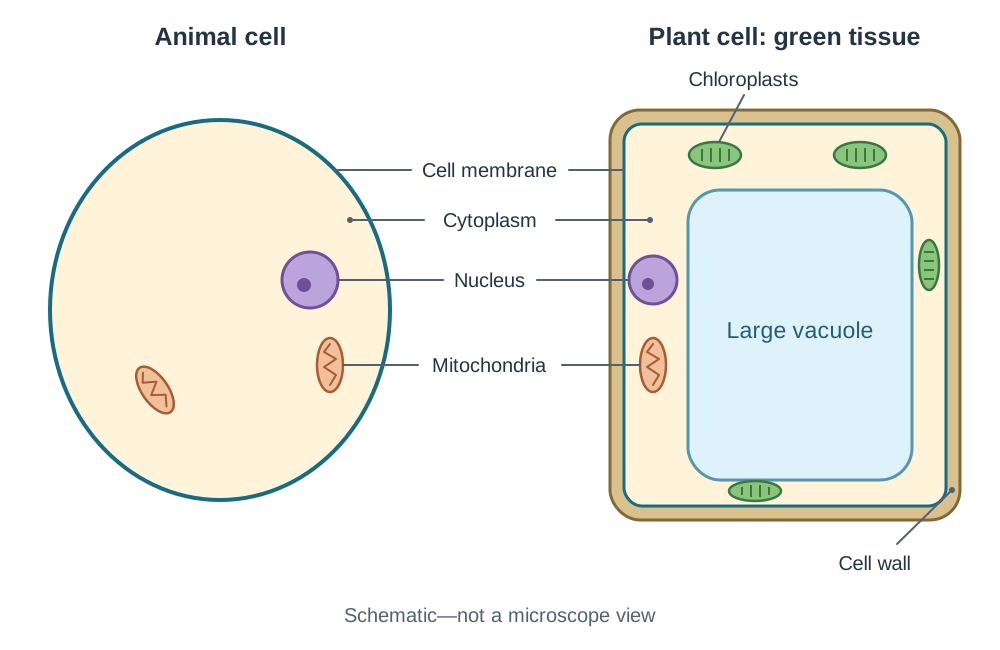
Onion bulb cells lack chloroplasts because the bulb grows underground in the dark away from light. They are still plant cells because they have a cellulose cell wall and a large vacuole.
Preparing Microscope Slides
Most cells are see-through, so a stain is added to give contrast and make parts such as the nucleus show up clearly.
General Method
- Place a thin sample on a clean glass slide in a drop of water.
- Add a drop of chemical stain.
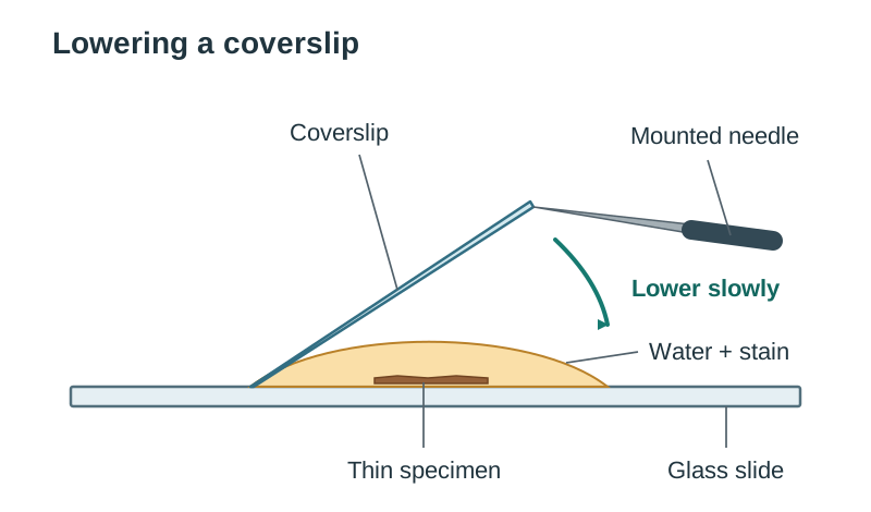
- Lower a coverslip slowly at an angle using a mounted needle to prevent trapping air bubbles.
Plant and Animal Specimens
- Plant tissue (onion epidermis): Peel a thin, transparent layer of onion skin with tweezers, place it on the slide, and stain it with iodine to highlight cell walls and nuclei.
- Animal tissue (cheek cells): Gently swab the inside of your cheek with a clean cotton bud (or use a prepared slide), smear it onto the slide, stain with methylene blue, and safely dispose of the swab.
Key terms
- Cell
- The basic building block of all living organisms.
- Unicellular organism
- A living thing made of only one cell, for example Euglena in pond water.
- Organelle
- A specialised structure within a cell that carries out a specific function.
- Cell membrane
- A thin layer around a cell that controls what enters and leaves.
- Cell wall
- A tough outer layer made of cellulose that provides shape and support to plant cells.
- Mitochondria
- Structures inside cells where respiration happens to release usable energy.
- Chloroplast
- A plant cell structure containing chlorophyll that absorbs light energy for photosynthesis.
- Stain
- A chemical dye, such as iodine or methylene blue, used to colour transparent cell parts under a microscope.
Check yourself
Name the three parts you would label on a diagram of an animal cell.
Nucleus, cytoplasm, and cell membrane.
What three structures are present in typical plant cells but absent in animal cells?
A cell wall, chloroplasts, and a large central vacuole.
Why do onion epidermal cells lack chloroplasts despite being plant cells?
Onion bulbs grow underground in the dark, so they do not receive light to carry out photosynthesis.
Which stains are used for onion cells and human cheek cells respectively?
Iodine solution for onion cells and methylene blue for cheek cells.
Why is a coverslip lowered slowly onto a slide at an angle?
To prevent air bubbles from becoming trapped over the specimen.